File:Accessory and cornuate navicular bone on dorsoplantar X-ray - annotated.jpg
Jump to navigation
Jump to search


Size of this preview: 800 × 373 pixels. Other resolutions: 320 × 149 pixels | 640 × 299 pixels | 1,200 × 560 pixels.
Original file (1,200 × 560 pixels, file size: 219 KB, MIME type: image/jpeg)
Summary
| Description |
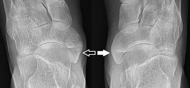
English: Dorsoplantar X-ray of the feet of a 44 year old woman with suspected pes cavus. The right foot (to the left in the image) has an accessory navicular bone (dark arrow), type 2. On the left foot (to the right in the image) it is fused with the navicular bone, forming a cornuate navicular bone (white arrow), which is type 3. |
| Date | |
| Source | Own work |
| Author | Mikael Häggström |
| Other versions |
 |
Licensing
I, the copyright holder of this work, hereby publish it under the following license:
| This file is made available under the Creative Commons CC0 1.0 Universal Public Domain Dedication. | |
| The person who associated a work with this deed has dedicated the work to the public domain by waiving all of their rights to the work worldwide under copyright law, including all related and neighboring rights, to the extent allowed by law. You can copy, modify, distribute and perform the work, even for commercial purposes, all without asking permission.
|
File history
Click on a date/time to view the file as it appeared at that time.
| Date/Time | Thumbnail | Dimensions | User | Comment | |
|---|---|---|---|---|---|
| current | 12:57, 30 October 2017 | | 1,200 × 560 (219 KB) | wikimediacommons>Mikael Häggström | User created page with UploadWizard |
File usage
There are no pages that use this file.