File:Ebstein1.jpg
Jump to navigation
Jump to search


Size of this preview: 800 × 341 pixels. Other resolutions: 320 × 136 pixels | 939 × 400 pixels.
Original file (939 × 400 pixels, file size: 60 KB, MIME type: image/jpeg)
Summary
| Description |
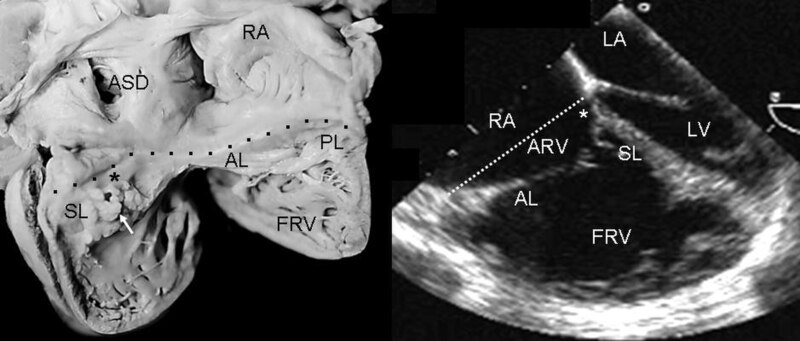
English: Internal view of the right chambers of a heart with Ebstein's anomaly with mild (Grade I) tethering of the septal leaflet (asterisk). Thickening of the free portion of the leaflet (arrow) is evident. The dotted line represents the atrioventricular junction where the tricuspid fibrous ring is located. The atrialized portion of the right ventricle is small. Note the patent foramen ovale type atrial septal defect. The 4 chamber echocardiographic image shows the same type of findings seen in the anatomic specimen. The dotted line indicates the plane of atrioventricular junction. The majority of the right ventricle is functional. Abbreviations: RA: Right atrium; ARV: Atrialized right ventricle; FRV: Functional right ventricle; AL: Anterior leaflet; PL: Posterior leaflet; SL: Septal leaflet; ASD: Atrial septal defect; LA: Left atrium; LV: Left ventricle. |
| Date | |
| Source | Muñoz-Castellanos L, Espinola-Zavaleta N, Kuri-Nivón M, Keirns C. Ebstein's Anomaly: anatomo-echocardiographic correlation. Cardiovasc Ultrasound. 5, 43. 2008. doi:10.1186/1476-7120-5-43. PMID 18034907. |
| Author | Luis Muñoz-Castellanos et al |
| Permission (Reusing this file) |
[1] |
Licensing
This file is licensed under the Creative Commons Attribution 2.0 Generic license.
- You are free:
- to share – to copy, distribute and transmit the work
- to remix – to adapt the work
- Under the following conditions:
- attribution – You must give appropriate credit, provide a link to the license, and indicate if changes were made. You may do so in any reasonable manner, but not in any way that suggests the licensor endorses you or your use.
File history
Click on a date/time to view the file as it appeared at that time.
| Date/Time | Thumbnail | Dimensions | User | Comment | |
|---|---|---|---|---|---|
| current | 18:55, 25 June 2008 | | 939 × 400 (60 KB) | wikimediacommons>Filip em | {{Information |Description={{en|1=Internal view of the right chambers of a heart with Ebstein's anomaly with mild (Grade I) tethering of the septal leaflet (asterisk). Thickening of the free portion of the leaflet (arrow) is evident. The dotted line repre |
File usage
There are no pages that use this file.