File:U fibres big.JPG

Original file (1,638 × 1,458 pixels, file size: 185 KB, MIME type: image/jpeg)
Summary
| Description | Desmyelinating Disorder |
| Date | |
| Source | Radiology picture of the day |
| Author | Dr. Laughlin Dawes |
| Permission (Reusing this file) |
Explanation
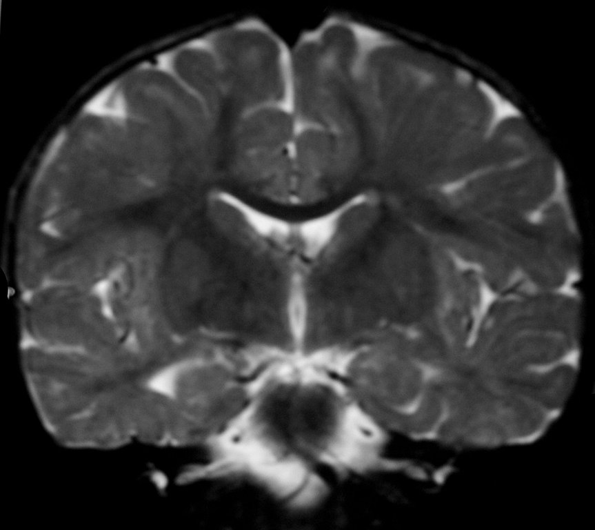
- "This 18-month old female presented with developmental delay. By this age myelination should be essentially
complete, yet there is residual T2 high signal in the subcortical white matter due to disruption of normal myelin formation. The differential diagnosis is depends on the head size.
With megalencephaly the possibilities are:
- Alexander's disease
- Canavan's disease
- van der Knapp’s leukodystrophy (but there is no cystic change)
Without megalencephaly: - in utero infection including Toxoplasma, Rubella, cytomegalovirus and herpes simplex virus. - mitochondrial cytopathy - Pelizeus-Merzbacher disease (but patient is female and PMD is X-linked recessive) - 18q deletion syndrome (the karyotype was normal) - Tuberous sclerosis (there were no other stigmata)
The more common leukodystrophies, metachromatic leukodystrophy and adrenoleukodystrophy, typically spare the subcortical U-fibres.
Credit: Dr Laughlin Dawes"Esta niña de 18 meses presenta retraso en el desarrollo. A esta edad la mielinización debería, en esencia, estar completa, aunque aún hay una alta señal de T2 en la materia blanca subcortical debido a la disrupción de la formación normal de mielina. El diagnóstico diferencial depende del volumen cefálico:
Con macrocefalia las posibilidades son:
- Enfermedad de alexander
- Enfermedad de Canavan
- Leucodistrofia de van der Knaap (sin cambios císticos)
Sin macrocefalia:
- Infecciones intrauterinas como toxoplasmosis, rubella, citomegalovirus y virus herpes simplex.
- Citopatía mitocondrial
- Enfermedad de Pelizeus-Merzbacher (Pero el paciente es niña y la enfermedad está ligada al X de forma recesiva)
- Sindrome de deleción del cromosoma 18q (El cariotipo era normal)
- Esclerosis tuberosa (no habia otras llagas)
Autor Dr Laughlin Dawes
Licensing
- You are free:
- to share – to copy, distribute and transmit the work
- to remix – to adapt the work
- Under the following conditions:
- attribution – You must give appropriate credit, provide a link to the license, and indicate if changes were made. You may do so in any reasonable manner, but not in any way that suggests the licensor endorses you or your use.
File history
Click on a date/time to view the file as it appeared at that time.
| Date/Time | Thumbnail | Dimensions | User | Comment | |
|---|---|---|---|---|---|
| current | 16:49, 10 March 2008 | | 1,638 × 1,458 (185 KB) | wikimediacommons>Gustavocarra | {{Information |Description=Desmyelinating Disorder |Source=[http://www.radpod.org/2006/11/19/dysmyelinating-disorder/ Radiology picture of the day] |Date=19/11/2006 |Author=Dr. Laughlin Dawes |Permission=Creative Commons |other_versions= }} ==Explanation |
File usage
There are no pages that use this file.