File:PMC3567970 1752-1947-7-35-1.png

From WikiMD.org
Jump to navigation Jump to search

PMC3567970_1752-1947-7-35-1.png(512 × 564 pixels, file size: 608 KB, MIME type: image/png)

This file is from Wikimedia Commons and may be used by other projects. The description on its file description page there is shown below.

Summary

Description
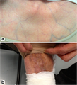
English: Skin findings. (a) Anterior chest wall: the skin was thin and subcutaneous veins were visible through it. (b) Back of the left foot: pigmented scars caused by cutaneous arteriovenous fistulae were visible.
Date
Source https://openi.nlm.nih.gov/detailedresult.php?img=PMC3567970_1752-1947-7-35-1&query=vascular+eds&lic=by&req=4&npos=14
Author Kashizaki F, Hatamochi A, Kamiya K, Yoshizu A, Okamoto H
Other versions

Licensing

w:en:Creative Commons
attribution
This file is licensed under the Creative Commons Attribution 2.0 Generic license.
You are free:
  • to share – to copy, distribute and transmit the work
  • to remix – to adapt the work
Under the following conditions:
  • attribution – You must give appropriate credit, provide a link to the license, and indicate if changes were made. You may do so in any reasonable manner, but not in any way that suggests the licensor endorses you or your use.
This file, which was originally posted to https://openi.nlm.nih.gov/detailedresult.php?img=PMC3567970_1752-1947-7-35-1&query=vascular+eds&lic=by&req=4&npos=14, was reviewed on 12 August 2019 by reviewer Explicit, who confirmed that it was available there under the stated license on that date.

Captions

Add a one-line explanation of what this file represents

Items portrayed in this file

depicts

File history

Click on a date/time to view the file as it appeared at that time.

Date/TimeThumbnailDimensionsUserComment
current15:32, 15 June 2018Thumbnail for version as of 15:32, 15 June 2018512 × 564 (608 KB)wikimediacommons>Steve MUser created page with UploadWizard

There are no pages that use this file.