File:12985 2016 678 Fig1 HTML (cropped).jpg
From WikiMD's WELLNESSPEDIA
Size of this preview: 515 × 600 pixels. Other resolution: 608 × 708 pixels.
Original file (608 × 708 pixels, file size: 87 KB, MIME type: image/jpeg)
Summary[edit]
| Summary | |
|---|---|
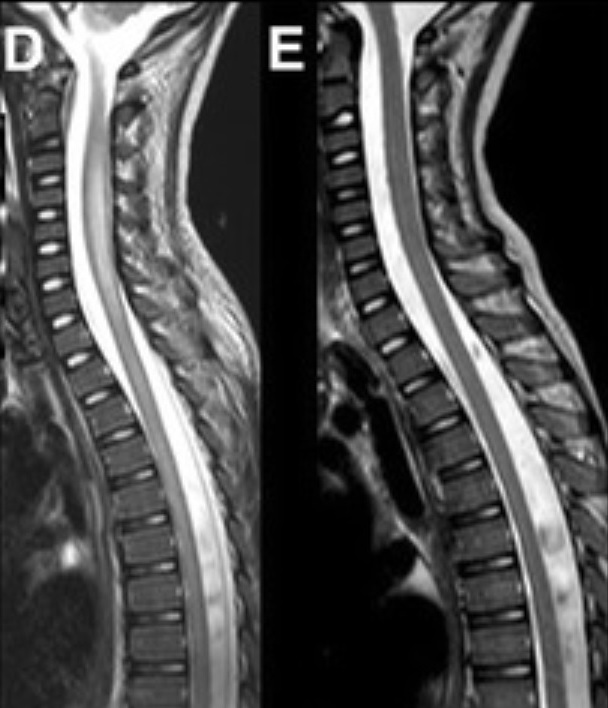
| Description | Magnetic resonance imaging (MRI) of the brain and spinal cord: a, axial T2 image at the level of the pons showing an increased signal in the tegmentum (white arrows), which was completely resolved in the follow up scan acquired 3 weeks later; b, sagittal and axial T2 image of the spinal cord (c, d) showing cord swelling, particularly at the cervical level (white, dot arrow), with extensive hyperintensity in the central cord (d), which was also completely resolved in the follow up images (e) acquired 3 weeks later |
| Source | Wikimedia Commons file page |
| Author | Susanna Esposito, Giovanna Chidini, Claudia Cinnante, Luisa Napolitano, Alberto Giannini, Leonardo Terranova, Hubert Niesters, Nicola Principi, and Edoardo Calderini |
| Permission | See original Commons license details. |
Licensing[edit]
Creative Commons Attribution 4.0 International (CC BY 4.0)
This file is licensed under the Creative Commons Attribution 4.0 International license.
You may share and adapt the material provided appropriate attribution is given.
Official license: CC BY 4.0
License page: CC BY 4.0
Original attribution and file history: Wikimedia Commons
File history
Click on a date/time to view the file as it appeared at that time.
| Date/Time | Thumbnail | Dimensions | User | Comment | |
|---|---|---|---|---|---|
| current | 02:10, 7 June 2026 | | 608 × 708 (87 KB) | Maintenance script (talk | contribs) | == Summary == Importing file |
You cannot overwrite this file.
File usage
The following file is a duplicate of this file (more details):
- File:12985 2016 678 Fig1 HTML (cropped).jpg from Wikimedia Commons
The following page uses this file:

.jpg&action=edit§ion=1)
.jpg)
.jpg&action=edit§ion=2)
.jpg)
.jpg&oldid=6598268)