File:20100916 011605 Mycelium.jpg
From WikiMD's WELLNESSPEDIA
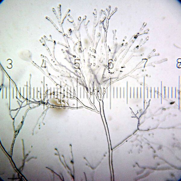
Size of this preview: 600 × 600 pixels. Other resolution: 1,600 × 1,600 pixels.
Original file (1,600 × 1,600 pixels, file size: 170 KB, MIME type: image/jpeg)
Summary[edit]
| Summary | |
|---|---|
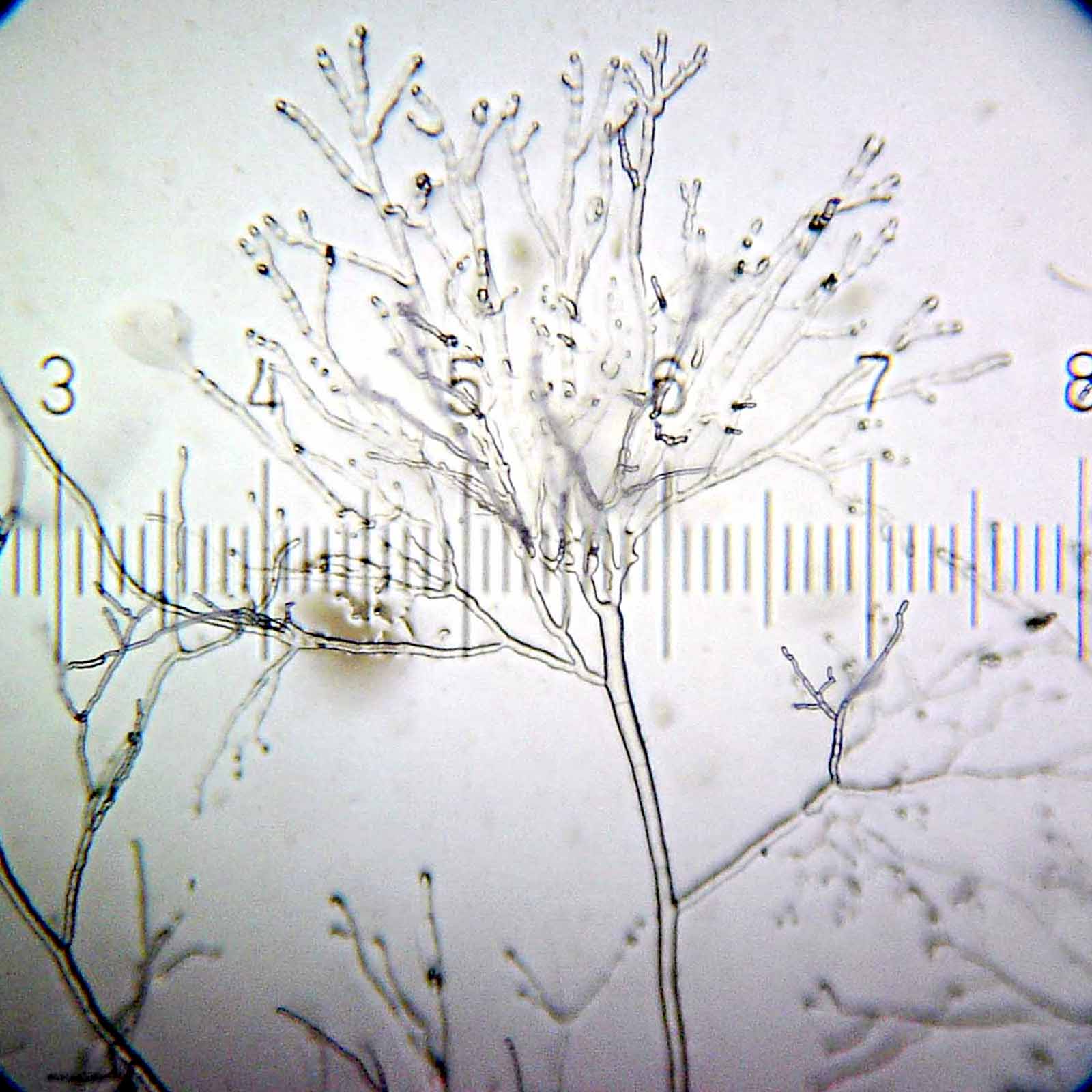
| Description | Early growth of mold from a bird dropping in EasyGel media in a Petri dish. At this point, all there is is the mycelium, and not the conidiophores that most people would recognize as “mold”. This eventually grew into a very impressive, fluffy white mold that filled the whole dish. 5× objective, 15× eyepiece; the numbered ticks are 230 µM apart. The scale of the full-sized version of this image is such that each pixel represents a 0.767 µM square. |
| Source | Wikimedia Commons file page |
| Author | Bob Blaylock |
| Permission | See original Commons license details. |
Licensing[edit]
Creative Commons Attribution-ShareAlike 3.0 Unported (CC BY-SA 3.0)
This file is licensed under the Creative Commons Attribution-ShareAlike 3.0 license.
Official license: CC BY-SA 3.0
License page: CC BY-SA 3.0
Original attribution and file history: Wikimedia Commons
File history
Click on a date/time to view the file as it appeared at that time.
| Date/Time | Thumbnail | Dimensions | User | Comment | |
|---|---|---|---|---|---|
| current | 02:06, 7 June 2026 | | 1,600 × 1,600 (170 KB) | Maintenance script (talk | contribs) | == Summary == Importing file |
You cannot overwrite this file.
File usage
The following page uses this file:
