File:2L6X Active Site Visualization E.Vitureira.png
From WikiMD's WELLNESSPEDIA

Size of this preview: 800 × 584 pixels. Other resolution: 1,054 × 770 pixels.
Original file (1,054 × 770 pixels, file size: 275 KB, MIME type: image/png)
Summary[edit]
| Summary | |
|---|---|
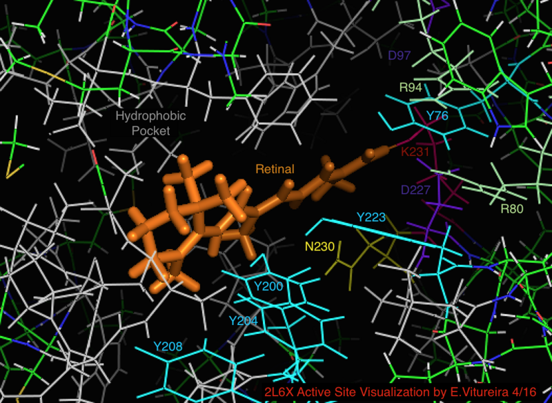
| Description | Visualization of the retinal bound active site of the 2L6X protein structure of pRhodopsin, residues color coded and labeled by activity, ligand is orange. |
| Source | Wikimedia Commons file page |
| Author | Elviture |
| Permission | See original Commons license details. |
Licensing[edit]
Creative Commons Attribution-ShareAlike 4.0 International (CC BY-SA 4.0)
This file is licensed under the Creative Commons Attribution-ShareAlike 4.0 International license.
You are free to:
- Share — copy and redistribute the material.
- Adapt — remix, transform, and build upon the material.
Under the following conditions:
- Attribution — appropriate credit must be given.
- ShareAlike — derivative works must be distributed under the same license.
Official license: CC BY-SA 4.0
License page: CC BY-SA 4.0
Original attribution and file history: Wikimedia Commons
File history
Click on a date/time to view the file as it appeared at that time.
| Date/Time | Thumbnail | Dimensions | User | Comment | |
|---|---|---|---|---|---|
| current | 02:14, 7 June 2026 | | 1,054 × 770 (275 KB) | Maintenance script (talk | contribs) | == Summary == Importing file |
You cannot overwrite this file.
File usage
The following page uses this file:
