File:3D-SIM-4 Anaphase 3 color.jpg
From WikiMD's WELLNESSPEDIA
Size of this preview: 790 × 600 pixels. Other resolution: 954 × 724 pixels.
Original file (954 × 724 pixels, file size: 264 KB, MIME type: image/jpeg)
Summary[edit]
| Summary | |
|---|---|
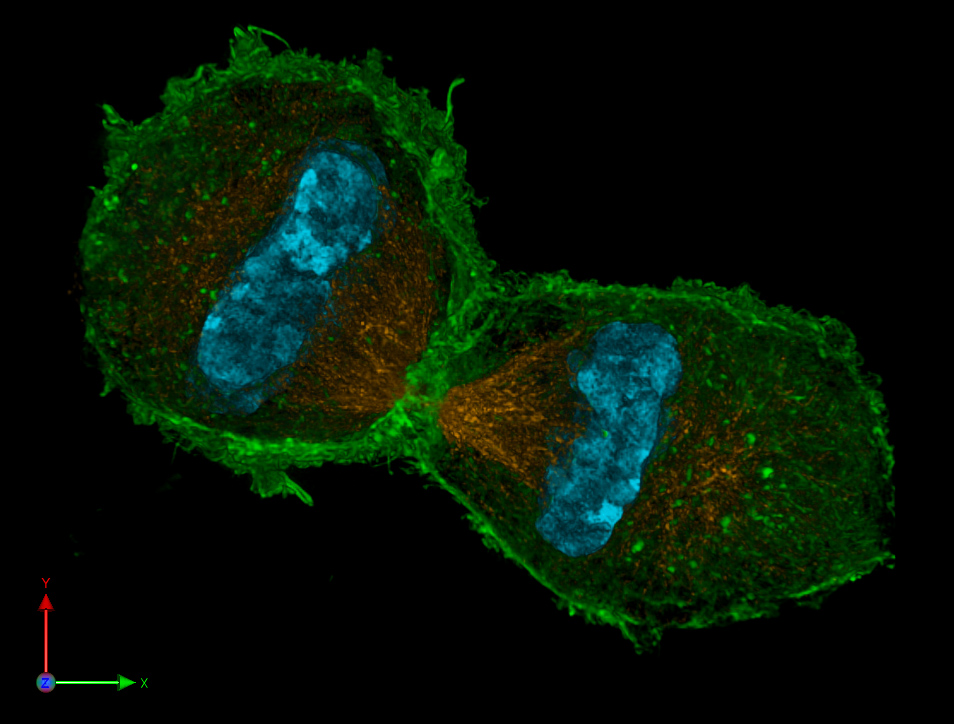
| Description | 3D representation of two mouse daughter nuclei in a late stage of nuclear division (Telophase). The spindle apparatus (anti-tubulin Immunostaining, orange), the actin cytoskeleton (Phalloidin staining, green) and chromatin (DAPI-staining, cyan) are visualized. For further information see: Schermelleh L, Carlton PM, Haase S, Shao L, Winoto L, Kner P, Burke B, Cardoso MC, Agard DA, Gustafsson MG, Leonhardt H, Sedat JW (June 2008). "Subdiffraction multicolor imaging of the nuclear periphery with 3D structured illumination microscopy". Science (journal) 320 (5881): 1332–6. DOI:10.1126/science.1156947. PMID 18535242. |
| Source | Wikimedia Commons file page |
| Author | Lothar Schermelleh |
| Permission | See original Commons license details. |
Licensing[edit]
Creative Commons Attribution-ShareAlike 3.0 Unported (CC BY-SA 3.0)
This file is licensed under the Creative Commons Attribution-ShareAlike 3.0 license.
Official license: CC BY-SA 3.0
License page: CC BY-SA 3.0
Original attribution and file history: Wikimedia Commons
File history
Click on a date/time to view the file as it appeared at that time.
| Date/Time | Thumbnail | Dimensions | User | Comment | |
|---|---|---|---|---|---|
| current | 02:11, 7 June 2026 | | 954 × 724 (264 KB) | Maintenance script (talk | contribs) | == Summary == Importing file |
You cannot overwrite this file.
File usage
The following file is a duplicate of this file (more details):
- File:3D-SIM-4 Anaphase 3 color.jpg from Wikimedia Commons
The following page uses this file:
