File:3D Whole Cell (3D-WC) model of a Mycoplasma genitalium cell.jpg
From WikiMD's WELLNESSPEDIA
Size of this preview: 793 × 599 pixels. Other resolution: 2,560 × 1,935 pixels.
Original file (2,560 × 1,935 pixels, file size: 829 KB, MIME type: image/jpeg)
Summary[edit]
| Summary | |
|---|---|
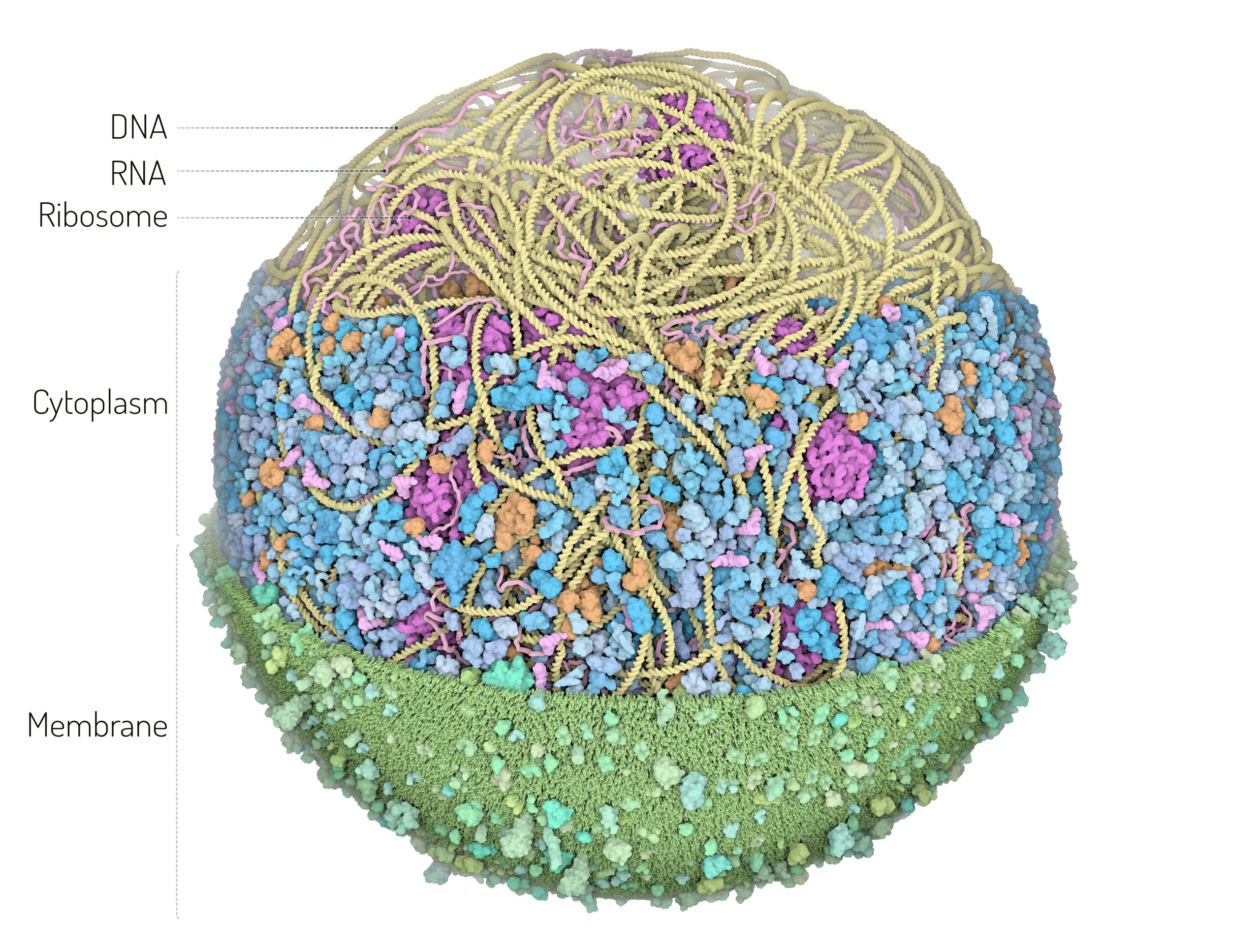
| Description | The 3D-WC model of Mycoplasma genitalium visualized with CellPACKgpu. Two clipping planes progressively hide parts of the model. The upper section highlights ribosomes (magenta), DNA (yellow) and mRNA (pink) filaments; the central section shows the bacterial nucleoid in the context of soluble macromolecules (DNA binding proteins in orange, cytoplasmic proteins in shades of blue, tRNAs in bright pink); the lower section shows the cell membrane (grey/green) with associated membrane proteins (shades of green). |
| Source | Wikimedia Commons file page |
| Author | Martina Maritan, Scripps Research |
| Permission | See original Commons license details. |
Licensing[edit]
Creative Commons Attribution 4.0 International (CC BY 4.0)
This file is licensed under the Creative Commons Attribution 4.0 International license.
You may share and adapt the material provided appropriate attribution is given.
Official license: CC BY 4.0
License page: CC BY 4.0
Original attribution and file history: Wikimedia Commons
File history
Click on a date/time to view the file as it appeared at that time.
| Date/Time | Thumbnail | Dimensions | User | Comment | |
|---|---|---|---|---|---|
| current | 02:11, 7 June 2026 | | 2,560 × 1,935 (829 KB) | Maintenance script (talk | contribs) | == Summary == Importing file |
You cannot overwrite this file.
File usage
The following file is a duplicate of this file (more details):
- File:3D Whole Cell (3D-WC) model of a Mycoplasma genitalium cell.jpg from Wikimedia Commons
The following page uses this file:

_model_of_a_Mycoplasma_genitalium_cell.jpg&action=edit§ion=1)
_model_of_a_Mycoplasma_genitalium_cell.jpg)
_model_of_a_Mycoplasma_genitalium_cell.jpg&action=edit§ion=2)
_model_of_a_Mycoplasma_genitalium_cell.jpg)
_model_of_a_Mycoplasma_genitalium_cell.jpg&oldid=6599969)