File:Bonthius1b.gif
From WikiMD's WELLNESSPEDIA

Size of this preview: 450 × 600 pixels. Other resolution: 600 × 800 pixels.
Original file (600 × 800 pixels, file size: 109 KB, MIME type: image/gif)
Summary[edit]
| Summary | |
|---|---|
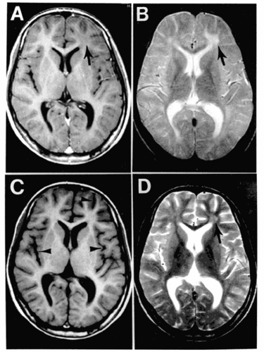
| Description | Subacute sclerosing panencephalitis. Figure 1. MRI scans of the brain at the time of presentation in the neurology clinic (A and B) and 3 months later (C and D). Panels A and C are T1-weighted images; B and D are T2-weighted images. The initial MRI scan (A and B) reveals a focal abnormality in the subcortical white matter of the left frontal lobe, consisting of a hypointense signal on the T1-weighted image (arrow in A) and a hyperintense signal on the T2-weighted image (arrow in B). In the followup scan, the focal abnormality in the left frontal lobe is less obvious than previously (arrow in D), but advanced and diffuse cortical atrophy is present, signified by the ventriculomegaly and markedly enlarged sulci (arrowheads in C). |
| Source | Wikimedia Commons file page |
| Author | Bonthius D, Stanek N, Grose C/ CDC |
| Permission | See original Commons license details. |
Licensing[edit]
Public Domain
This file is in the public domain and may be used without restriction.
Please see the linked source page for the original file history, attribution information, and licensing details.
Original attribution and file history: Wikimedia Commons
File history
Click on a date/time to view the file as it appeared at that time.
| Date/Time | Thumbnail | Dimensions | User | Comment | |
|---|---|---|---|---|---|
| current | 13:40, 8 June 2026 | | 600 × 800 (109 KB) | Maintenance script (talk | contribs) | == Summary == Importing file |
You cannot overwrite this file.
File usage
The following page uses this file:
