File:Cardiovasc Ultrasound LVNC 3.jpg
From WikiMD's WELLNESSPEDIA
No higher resolution available.
Cardiovasc_Ultrasound_LVNC_3.jpg (400 × 240 pixels, file size: 21 KB, MIME type: image/jpeg)
Summary[edit]
| Summary | |
|---|---|
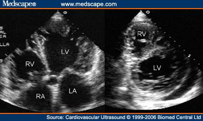
| Description | Transthoracic two-dimensional echocardiogram in apical four chamber and parasternal short axis at the level of both ventricles demonstrate dilatation, deep trabeculae and intertrabecular recesses in the inferior, lateral, anterior walls, middle and apical portions of the septum and apex of the left ventricle. The right ventricle also shows evidence of noncompaction. A posterolateral pericardial effusion is also present. Others abbreviations as before.
Non-compacted Cardiomyopathy: Clinical-Echocardiographic Study by Nilda Espinola-Zavaleta; M. Elena Soto; Luis Muñóz Castellanos; Silvio Játiva-Chávez; Candace Keirns in March 2007 by www.medscape.com as stated in the article Cardiovasc Ultrasound. 2006;4(1) ©2006 Espinola-Zavaleta et al; licensee BioMed Central Ltd. This is an Open Access article distributed under the terms of the Creative Commons Attribution License (https://creativecommons.org/licenses/by/2.0), which permits unrestricted use, distribution, and reproduction in any medium, provided the original work is properly cited. |
| Source | Wikimedia Commons file page |
| Author | The original uploader was Dexcel at English Wikipedia. |
| Permission | See original Commons license details. |
Licensing[edit]
License: CC BY 2.5
License page: CC BY 2.5
Original attribution and file history: Wikimedia Commons
File history
Click on a date/time to view the file as it appeared at that time.
| Date/Time | Thumbnail | Dimensions | User | Comment | |
|---|---|---|---|---|---|
| current | 13:39, 8 June 2026 | | 400 × 240 (21 KB) | Maintenance script (talk | contribs) | == Summary == Importing file |
You cannot overwrite this file.
File usage
The following page uses this file:
