File:Cerebral MRI of Taenia Crassiceps tapeworm infection.jpg
From WikiMD's WELLNESSPEDIA
No higher resolution available.
Cerebral_MRI_of_Taenia_Crassiceps_tapeworm_infection.jpg (600 × 404 pixels, file size: 33 KB, MIME type: image/jpeg)
Summary[edit]
| Summary | |
|---|---|
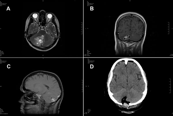
| Description | Magnetic resonance (MR) and computed tomographic images of the brain of a 51-year-old woman infected with Taenia crassiceps tapeworm larvae, Germany. A) Transverse view, T1-weighted MR image. The 30 × 30 mm parasitic lesion with perifocal edema is located in the right hemisphere of the cerebellum and caused ataxia, headache, and nausea. The fourth ventricle is compressed. B) Coronal view, T2-weighted MR image. The cyst-like appearance of the parasitic tissue is clearly visible. This lesion can be misinterpreted as cerebral echinococcosis, racemose cysticercosis caused by a Taenia solium tapeworm, or coenurosis. C) Sagittal view, MR image with contrast enhancing agent. D) Transverse view, computed tomographic image after surgery. No residual parasitic masses, only the parenchymal defect in the cerebellum after resection of T. crassiceps tapeworm larvae, are visible. |
| Source | Wikimedia Commons file page |
| Author | Vasileios Ntoukas, Dennis Tappe, Daniel Pfütze, Michaela Simon, and Thomas Holzmann |
| Permission | See original Commons license details. |
Licensing[edit]
Public Domain
This file is in the public domain and may be used without restriction.
Please see the linked source page for the original file history, attribution information, and licensing details.
Original attribution and file history: Wikimedia Commons
File history
Click on a date/time to view the file as it appeared at that time.
| Date/Time | Thumbnail | Dimensions | User | Comment | |
|---|---|---|---|---|---|
| current | 13:40, 8 June 2026 | | 600 × 404 (33 KB) | Maintenance script (talk | contribs) | == Summary == Importing file |
You cannot overwrite this file.
File usage
The following page uses this file:
