File:Ferruginous body.jpg
From WikiMD's WELLNESSPEDIA
Size of this preview: 529 × 599 pixels. Other resolution: 2,512 × 2,844 pixels.
Original file (2,512 × 2,844 pixels, file size: 2.1 MB, MIME type: image/jpeg)
Summary[edit]
| Summary | |
|---|---|
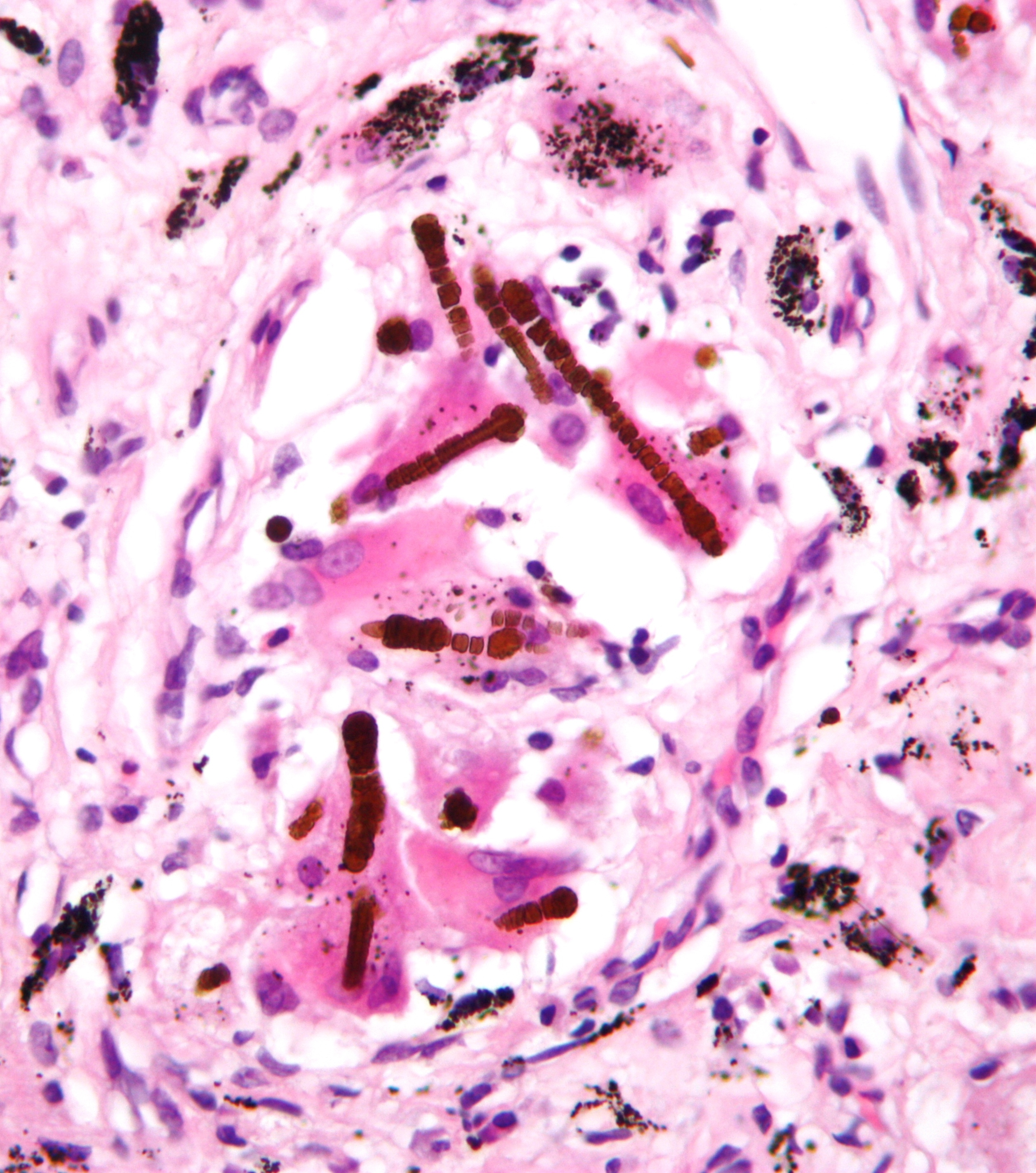
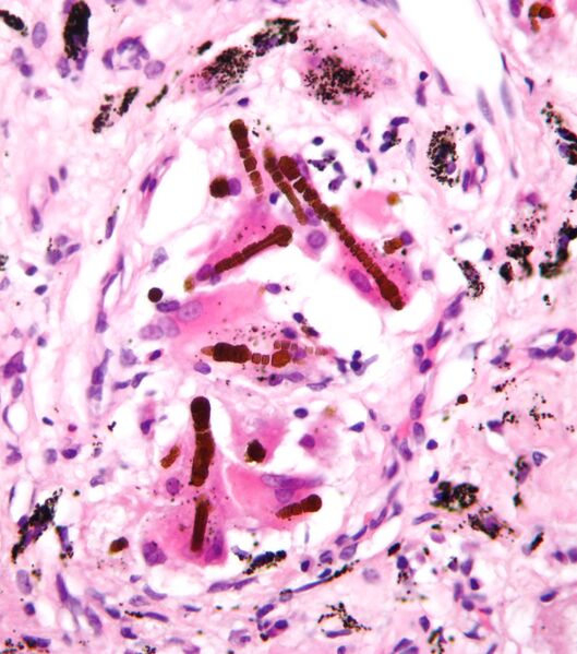
| Description | High magnification micrograph of ferruginous bodies, also asbestos bodies, as seen in asbestosis. Lung biopsy. H&E stain. Ferrunginous bodies are rust coloured (red/brown) baton-shaped segmented rods.
Anthracotic pigment (clumps of small black particles) is also seen on the micrograph (at the periphery). See also Image:Asbestosis high mag.jpg - Ferruginous bodies in asbestosis, same case - lower magnification. |
| Source | Wikimedia Commons |
| Author | Nephron |
| Permission | See Commons |
Licensing[edit]
Creative Commons Attribution-ShareAlike 3.0 Unported (CC BY-SA 3.0)
This file is licensed under the Creative Commons Attribution-ShareAlike 3.0 license.
Official license: CC BY-SA 3.0
Original attribution and file history: Wikimedia Commons
File history
Click on a date/time to view the file as it appeared at that time.
| Date/Time | Thumbnail | Dimensions | User | Comment | |
|---|---|---|---|---|---|
| current | 01:21, 2 June 2026 | | 2,512 × 2,844 (2.1 MB) | Maintenance script (talk | contribs) | == Summary == Importing file |
You cannot overwrite this file.
File usage
The following 2 pages use this file:
