File:Fibrothorax chest x-ray.jpg
From WikiMD's medical encyclopedia
Size of this preview: 609 × 600 pixels. Other resolutions: 244 × 240 pixels | 487 × 480 pixels | 780 × 768 pixels | 1,195 × 1,177 pixels.
Original file (1,195 × 1,177 pixels, file size: 240 KB, MIME type: image/jpeg)
Summary
| Description |
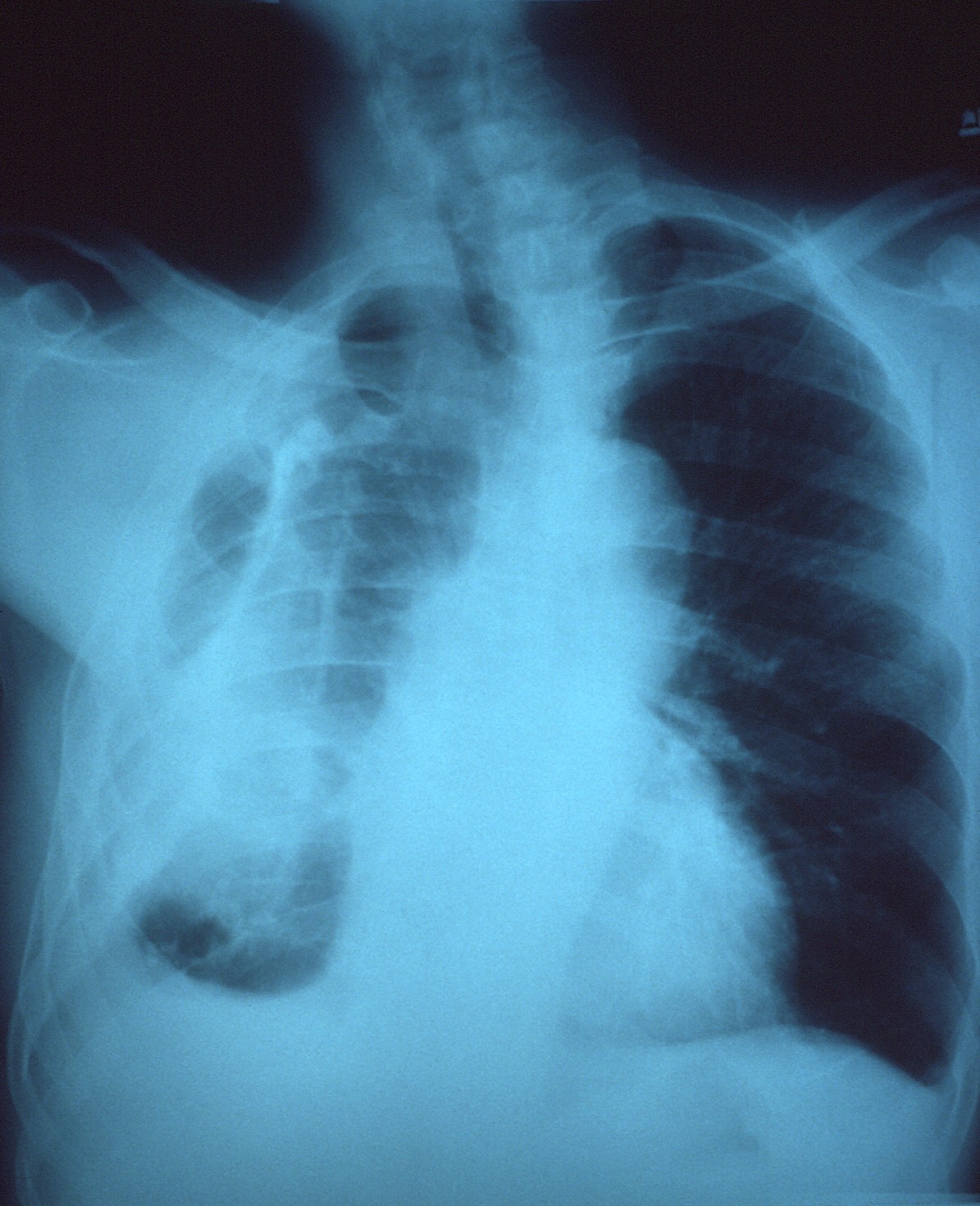
English: This AP chest x-ray revealed the presence of a right fibrothorax, which was thought to be due to a previous empyema.
An empyema, or pus-filled cavity located between the two pleural membranes, i.e., parietal and visceral pleurae, can give rise to fibrotic changes as the infection resolves, much like a scar formation, which appears as a denser area on x-ray. |
| Source | Public Health Image Library (PHIL), Image 6243 |
| Author | CDC/ Dr. Thomas Hooten |
Licensing
This file is a work of the Centers for Disease Control and Prevention, part of the United States Department of Health and Human Services, taken or made as part of an employee's official duties. As a work of the U.S. federal government, the file is in the public domain.
|
|
This media comes from the Centers for Disease Control and Prevention's Public Health Image Library (PHIL), with identification number #6243. Note: Not all PHIL images are public domain; be sure to check copyright status and credit authors and content providers.
|
File history
Click on a date/time to view the file as it appeared at that time.
| Date/Time | Thumbnail | Dimensions | User | Comment | |
|---|---|---|---|---|---|
| current | 10:47, 16 March 2025 | | 1,195 × 1,177 (240 KB) | Doc James | Cropped 20 % vertically, 20 % areawise using CropTool with precise mode. |
File usage
The following page uses this file: