File:GFAP gliosis.jpg
From WikiMD's WELLNESSPEDIA
Size of this preview: 800 × 312 pixels. Other resolution: 1,859 × 724 pixels.
Original file (1,859 × 724 pixels, file size: 598 KB, MIME type: image/jpeg)
Summary[edit]
| Summary | |
|---|---|
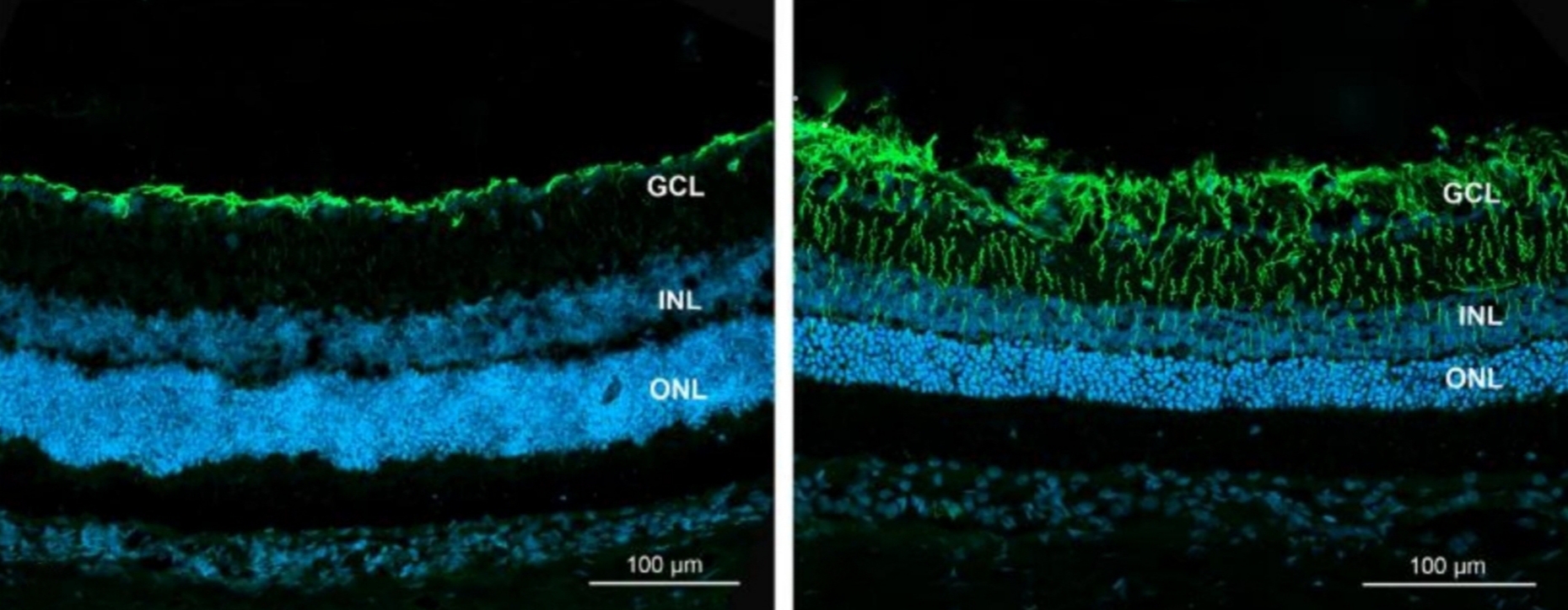
| Description | Confocal microscopy imaging of rat retinal sections immunolabeled for glial fibrillary acidic protein (GFAP) reveals distinct morphological changes in Müller cell processes. In normal retinas on the left, GFAP expression is predominantly localized to the innermost layers, namely the nerve fiber layer and the ganglion cell layer (GCL). However, on the right, there is a marked increase in GFAP-positive fibers, indicating pronounced hypertrophy and thickening of Müller cell processes. These hypertrophic processes extended through the inner nuclear layer (INL) and outer nuclear layer (ONL), strongly indicative of retinal gliosis (Nuclei, stained with DAPI, appear blue under microscopy). |
| Source | Wikimedia Commons file page |
| Author | Ziółkowska N, Lewczuk B, Szyryńska N, Rawicka A, Vyniarska A |
| Permission | See original Commons license details. |
Licensing[edit]
Creative Commons Attribution 4.0 International (CC BY 4.0)
This file is licensed under the Creative Commons Attribution 4.0 International license.
You may share and adapt the material provided appropriate attribution is given.
Official license: CC BY 4.0
License page: CC BY 4.0
Original attribution and file history: Wikimedia Commons
File history
Click on a date/time to view the file as it appeared at that time.
| Date/Time | Thumbnail | Dimensions | User | Comment | |
|---|---|---|---|---|---|
| current | 13:37, 8 June 2026 | 1,859 × 724 (598 KB) | Maintenance script (talk | contribs) | == Summary == Importing file |
You cannot overwrite this file.
File usage
The following file is a duplicate of this file (more details):
- File:GFAP gliosis.jpg from Wikimedia Commons
The following page uses this file:
