File:Gallbladder cholesterolosis intermed mag cropped.jpg

Original file (1,740 × 1,312 pixels, file size: 1.17 MB, MIME type: image/jpeg)
Summary[edit]
| Summary | |
|---|---|
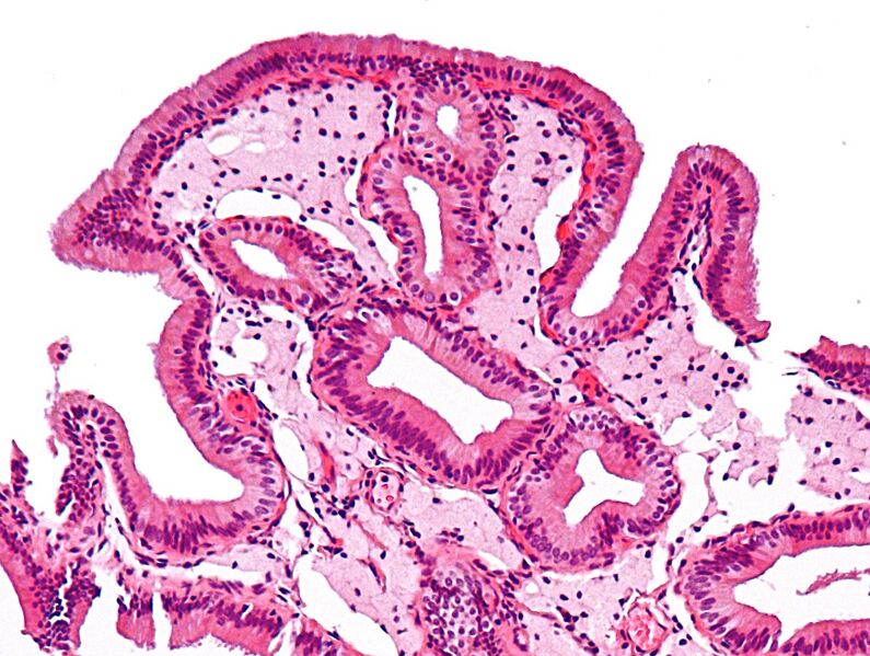
| Description | Intermediate magnification micrograph of cholesterolosis of the gallbladder. Cholecystectomy specimen. H&E stain.
العربية: صورة مجهرية لكولسترولية المرارة، وهو مرض شائع جدًا في المرارة. Dead lipid laden macrophages (foam cells) are seen in the finger-like projections into the gallbladder lumen. It should be apparent that this is gallbladder, as no muscularis mucosae is present (as elsewhere in the gastrointestinal tract). The blood vessels are congested and the subserosa edematous. Cholesterolosis is often associated with cholecystitis. See also Image:Gallbladder cholesterolosis low mag.jpg Image:Gallbladder cholesterolosis intermed mag.jpg - uncropped version of this image Another case: Image:Gallbladder cholesterolosis micro.jpg |
| Source | Wikimedia Commons file page |
| Author | Nephron |
| Permission | See original Commons license details. |
Licensing[edit]
Creative Commons Attribution-ShareAlike 3.0 Unported (CC BY-SA 3.0)
This file is licensed under the Creative Commons Attribution-ShareAlike 3.0 license.
Official license: CC BY-SA 3.0
Original attribution and file history: Wikimedia Commons
File history
Click on a date/time to view the file as it appeared at that time.
| Date/Time | Thumbnail | Dimensions | User | Comment | |
|---|---|---|---|---|---|
| current | 12:50, 29 May 2026 | | 1,740 × 1,312 (1.17 MB) | Maintenance script (talk | contribs) | == Summary == Importing file |
You cannot overwrite this file.
File usage
The following 2 pages use this file:
