File:Gastrointestinal Stromal Tumor (GIST) of Stomach.jpg
Gastrointestinal_Stromal_Tumor_(GIST)_of_Stomach.jpg (650 × 448 pixels, file size: 73 KB, MIME type: image/jpeg)
Summary[edit]
| Summary | |
|---|---|
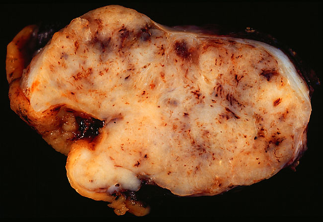
| Description | Gastrointestinal Stromal Tumor of Stomach
This 6.5-cm tumor was removed from the stomach of a 68-year-old man who presented with gastric bleeding. The lesion was first noted by upper GI endoscopy, which showed a deep bleeding ulcer thought to represent a vascular malformation. At laparatomy, examination of the external surface of the stomach showed this oblong tumor that extended from the gastric wall superiorly to the surface of the liver, to which the tumor was stuck. The surgeon dissected the tumor off the liver and included a uniform rim of normal hepatic tissue to obtain a clear margin. He also made sure to include a circumferential skirt of normal gastric wall with the resection. The diagnosis of gastrointestinal stromal tumor was made intraoperatively by frozen section. With this information, and with pathologic evaluation to assess margins, the surgeon was able to avoid doing a total gastrectomy. On permanent section, the tumor showed uniform, wispy spindle cells with no mitotic activity. Although one can't be sure even a bland GIST won't recur, the rate of recurrence is low in gastric tumors with low mitotic counts. Published experience has shown that adherence of the tumor to the liver is not an adverse prognostic influence. On the other hand, a tumor size greater than 5 cm has negative prognostic implications. Because of their unpredictable behavior, all GIST cases need to be followed indefinitely. Surgical resection of metastases can be beneficial, and now the availability of STI571 (Gleevec), a specific inhibitor of the tyrosine kinase produced by the GIST's mutant c-kit proto-oncogene, means that recurrences can be treated with specific chemotherapy. In this photo, the gastric wall with the ulcer is on the left side of the image. Note how well the mucosa is demarcated from the tumor surface. Tags of dark liver tissue, barely visible, adhere to the tumor on the right side of the image. This photo was shot with a Minolta X-370 with 100mm bellows lens on Ektachrome Elite 100 daylight film, through a blue filter to correct for tungsten illumination. The film image was scanned on a Polaroid SprintScan using a Power Macintosh 7100/66AV and edited with Adobe Photoshop version 3.04. Editing included sharpening with the Unsharp Mask command, enhancing contrast with the Levels command, and adjusting the image size to fit to the Web page. The edited image was saved in JPEG format with compression set at "High" quality. Photograph by Ed Uthman, MD. Public domain. Posted 22 Aug 01 |
| Source | Wikimedia Commons file page |
| Author | See Wikimedia Commons file page |
| Permission | See original Commons license details. |
Licensing[edit]
Public Domain
This file is in the public domain and may be used without restriction.
Please see the linked source page for the original file history, attribution information, and licensing details.
Original attribution and file history: Wikimedia Commons
File history
Click on a date/time to view the file as it appeared at that time.
| Date/Time | Thumbnail | Dimensions | User | Comment | |
|---|---|---|---|---|---|
| current | 13:37, 8 June 2026 | | 650 × 448 (73 KB) | Maintenance script (talk | contribs) | == Summary == Importing file |
You cannot overwrite this file.
File usage
The following page uses this file:

_of_Stomach.jpg&action=edit§ion=1)
_of_Stomach.jpg)
_of_Stomach.jpg&action=edit§ion=2)
_of_Stomach.jpg&oldid=6604178)