File:Histopathology of a blood clot with postmortem bacterial growth.jpg
From WikiMD's WELLNESSPEDIA
Size of this preview: 715 × 600 pixels. Other resolution: 731 × 613 pixels.
Original file (731 × 613 pixels, file size: 150 KB, MIME type: image/jpeg)
Summary[edit]
| Summary | |
|---|---|
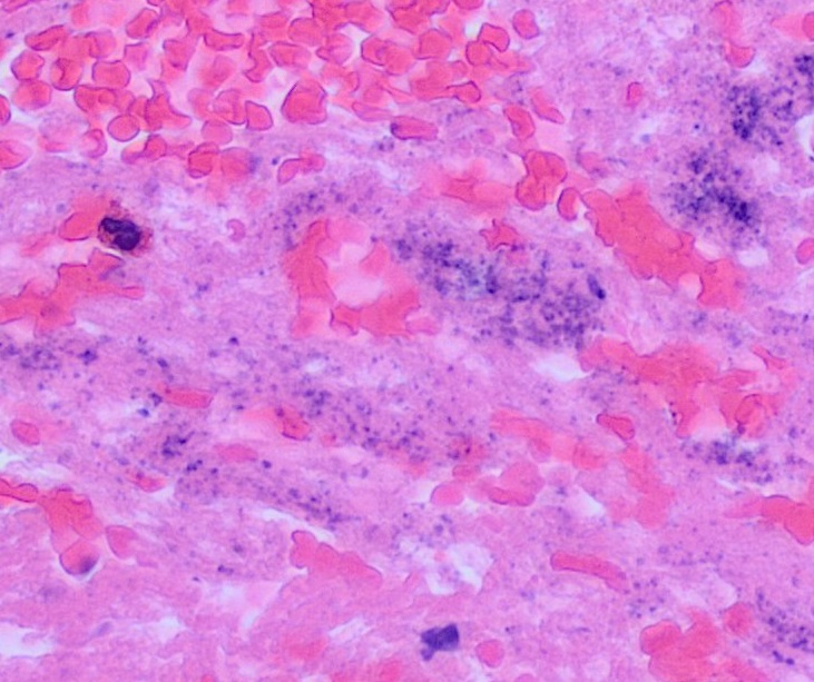
| Description | Histopathology from an autopsy, of a blood clot with bacterial growth in the fibrin, seen as basophilic and equally sized tiny granules on H&E stain. The neutrophils (one seen at left) are not increased compared to what is normally seen in blood. Hence, both the clot and bacterial growth are consistent with postmortem changes. |
| Source | Wikimedia Commons file page |
| Author | Mikael Häggström, M.D. Author info - Reusing images- Conflicts of interest: None Mikael Häggström, M.D.Consent note: Consent from the patient or patient's relatives is regarded as redundant, because of absence of identifiable features (List of HIPAA identifiers) in the media and case information (See also HIPAA case reports guidance). |
| Permission | See original Commons license details. |
Licensing[edit]
License: CC0
License page: CC0
Original attribution and file history: Wikimedia Commons
File history
Click on a date/time to view the file as it appeared at that time.
| Date/Time | Thumbnail | Dimensions | User | Comment | |
|---|---|---|---|---|---|
| current | 22:34, 8 June 2026 | | 731 × 613 (150 KB) | Maintenance script (talk | contribs) | == Summary == Importing file |
You cannot overwrite this file.
File usage
The following page uses this file:
