File:Histopathology of siderophage in chronic pulmonary congestion.jpg
From WikiMD's WELLNESSPEDIA
Size of this preview: 636 × 600 pixels. Other resolution: 883 × 833 pixels.
Original file (883 × 833 pixels, file size: 115 KB, MIME type: image/jpeg)
Summary[edit]
| Summary | |
|---|---|
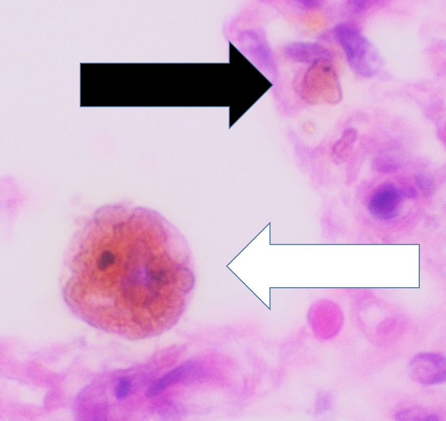
| Description | Histopathology chronic pulmonary congestion, showing a siderophage (white arrow, characterized by coarse brown pigment, which is slightly refractile), and interstitium with edema, hemosiderin deposition (black arrow) and collagenous thickening. H&E stain. |
| Source | Wikimedia Commons file page |
| Author | Mikael Häggström, M.D. Author info - Reusing images- Conflicts of interest: None Mikael Häggström, M.D.Consent note: Consent from the patient or patient's relatives is regarded as redundant, because of absence of identifiable features (List of HIPAA identifiers) in the media and case information (See also HIPAA case reports guidance). |
| Permission | See original Commons license details. |
Licensing[edit]
License: CC0
License page: CC0
Original attribution and file history: Wikimedia Commons
File history
Click on a date/time to view the file as it appeared at that time.
| Date/Time | Thumbnail | Dimensions | User | Comment | |
|---|---|---|---|---|---|
| current | 03:18, 5 June 2026 | | 883 × 833 (115 KB) | Maintenance script (talk | contribs) | == Summary == Importing file |
You cannot overwrite this file.
File usage
The following 2 pages use this file:
