File:Human motor map.jpg
From WikiMD's WELLNESSPEDIA
Size of this preview: 786 × 600 pixels. Other resolution: 1,214 × 926 pixels.
Original file (1,214 × 926 pixels, file size: 255 KB, MIME type: image/jpeg)
Summary[edit]
| Summary | |
|---|---|
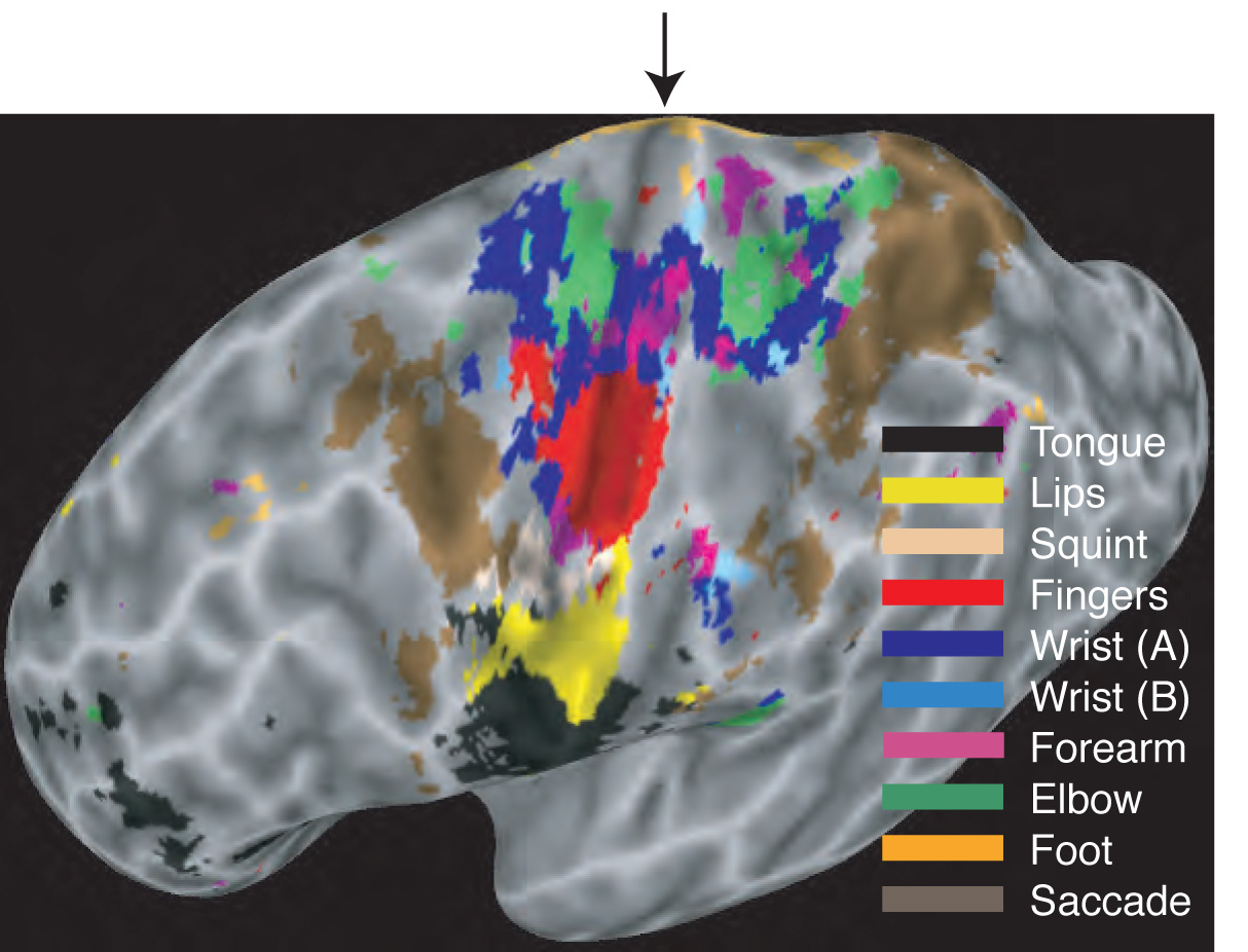
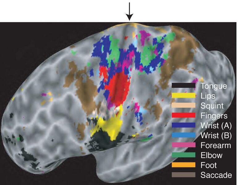
| Description | Map of the body in the human brain. The cerebral cortex has been “inflated” to show the results more clearly. Movements of different body parts evoked activity in the primary motor cortex (left of the central sulcus, shown by the arrow) and primary somatosensory cortex (right of the central sulcus). The map also shows eye-movement activity in the frontal eye field and the parietal cortex. The map is a winner-take-all. Although different body parts activated overlapping areas of cortex, only the strongest activations are indicated. The map in the primary motor cortex contains two common complexities. First, the hand representation (red) is surrounded on three sides by a representation of the wrist. Second, there are two distinct hand representations possibly corresponding to areas 4a and 4p. |
| Source | Wikimedia Commons file page |
| Author | Michael Graziano |
| Permission | See original Commons license details. |
Licensing[edit]
License: CC BY-SA 1.0
License page: CC BY-SA 1.0
Original attribution and file history: Wikimedia Commons
File history
Click on a date/time to view the file as it appeared at that time.
| Date/Time | Thumbnail | Dimensions | User | Comment | |
|---|---|---|---|---|---|
| current | 03:33, 5 June 2026 | | 1,214 × 926 (255 KB) | Maintenance script (talk | contribs) | == Summary == Importing file |
You cannot overwrite this file.
File usage
The following 2 pages use this file:
