File:Lectures on appendicitis and notes on other subjects (1897) (14764898745).jpg
Original file (1,208 × 2,468 pixels, file size: 330 KB, MIME type: image/jpeg)
Summary[edit]
| Summary | |
|---|---|
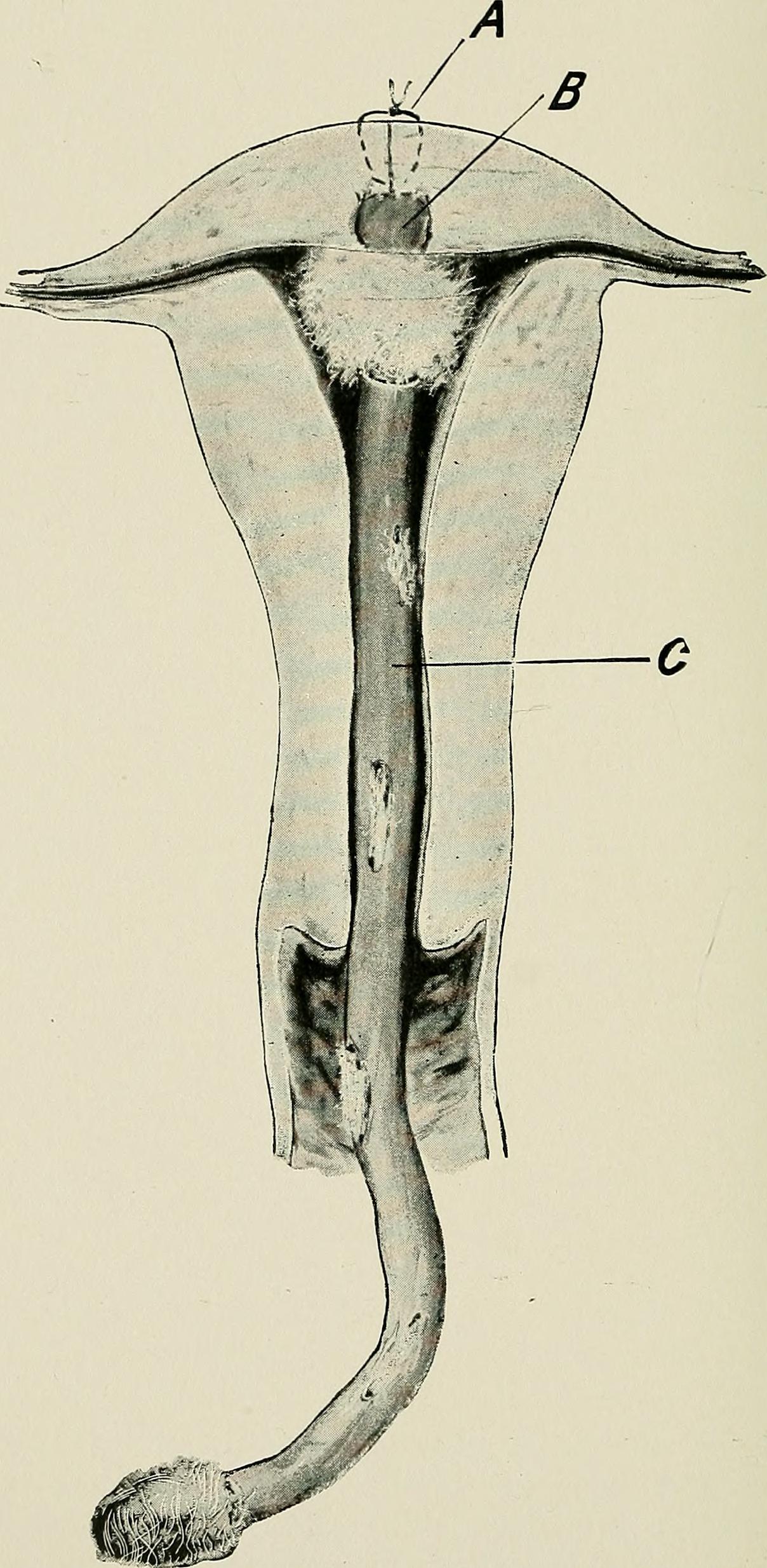
| Description | Placement of an ovarian graft
Identifier: lecturesonappend1897morr (find matches) Title: Lectures on appendicitis and notes on other subjects Year: 1897 (1890s) Authors: Morris, Robert T. (Robert Tuttle), 1857-1945 Subjects: Appendicitis Surgery, Operative Publisher: New York, G.P. Putnam's Sons Contributing Library: Columbia University Libraries Digitizing Sponsor: Open Knowledge Commons View Book Page: Book Viewer About This Book: Catalog Entry View All Images: All Images From Book Click here to view book online to see this illustration in context in a browseable online version of this book. Text Appearing Before Image: , and is fastened in place by a fine cat-gut suture that serves at the same time to partially close the slitin the uterus. Other sutures that are necessary for closing thewound are introduced, and a drainage wick of gauze is placed inthe uterine canal leading out through the vagina into a receivingmass of gauze at the vulva. The fundus of the uterus that is toreceive a graft is reached by way of an anterior abdominal inci-sion, or preferably by way of the vagina through a button-holeopening into Douglas ciil de sac. The fundus is readily turneddown into the vagina, and after receiving the graft is turned backinto the abdomen again, and the patient is then ready to get outof bed in two or three days. The gauze drain from the uterus isremoved at the end of forty-eight hours after the operation andthe case should require little further treatment. i=;S Notes. In cases in which the oviduct is chosen as the place for insert-ing an ovarian graft, it is difficult to find the lumen of the tube Text Appearing After Image: Fig. 66.—A.—Suture of slit through which graft was inserted.B.—Ovarian graft.C.—Drainage wick. Ovarian Transplantation. 159 if the latter has been cut short, because the muscular sheath con-tracts and inverts margins of the mucous tube. Before attempt-ing to insert the graft in such a case it is best to pass a probethrough the lumen of the oviduct into the uterus first and thenamputate the oviduct about the probe, suturing mucosa andperitoneum together at any one point in the circular cut beforecompleting the division. This will prevent inversion of mucosawhen the muscularis contracts and will allow us to keep the graft Note About Images Please note that these images are extracted from scanned page images that may have been digitally enhanced for readability - coloration and appearance of these illustrations may not perfectly resemble the original work. |
| Source | Wikimedia Commons file page |
| Author | Internet Archive Book Images |
| Permission | See original Commons license details. |
Licensing[edit]
License: No restrictions
License page: No restrictions
Original attribution and file history: Wikimedia Commons
File history
Click on a date/time to view the file as it appeared at that time.
| Date/Time | Thumbnail | Dimensions | User | Comment | |
|---|---|---|---|---|---|
| current | 22:26, 8 June 2026 | | 1,208 × 2,468 (330 KB) | Maintenance script (talk | contribs) | == Summary == Importing file |
You cannot overwrite this file.
File usage
The following file is a duplicate of this file (more details):
- File:Lectures on appendicitis and notes on other subjects (1897) (14764898745).jpg from Wikimedia Commons
The following page uses this file:

_(14764898745).jpg&action=edit§ion=1)
_(14764898745).jpg)
_(14764898745).jpg&action=edit§ion=2)
_(14764898745).jpg)
_(14764898745).jpg&oldid=6609721)