File:Mixed germ cell tumour - very high mag.jpg
Original file (4,272 × 2,848 pixels, file size: 4.38 MB, MIME type: image/jpeg)
Summary[edit]
| Summary | |
|---|---|
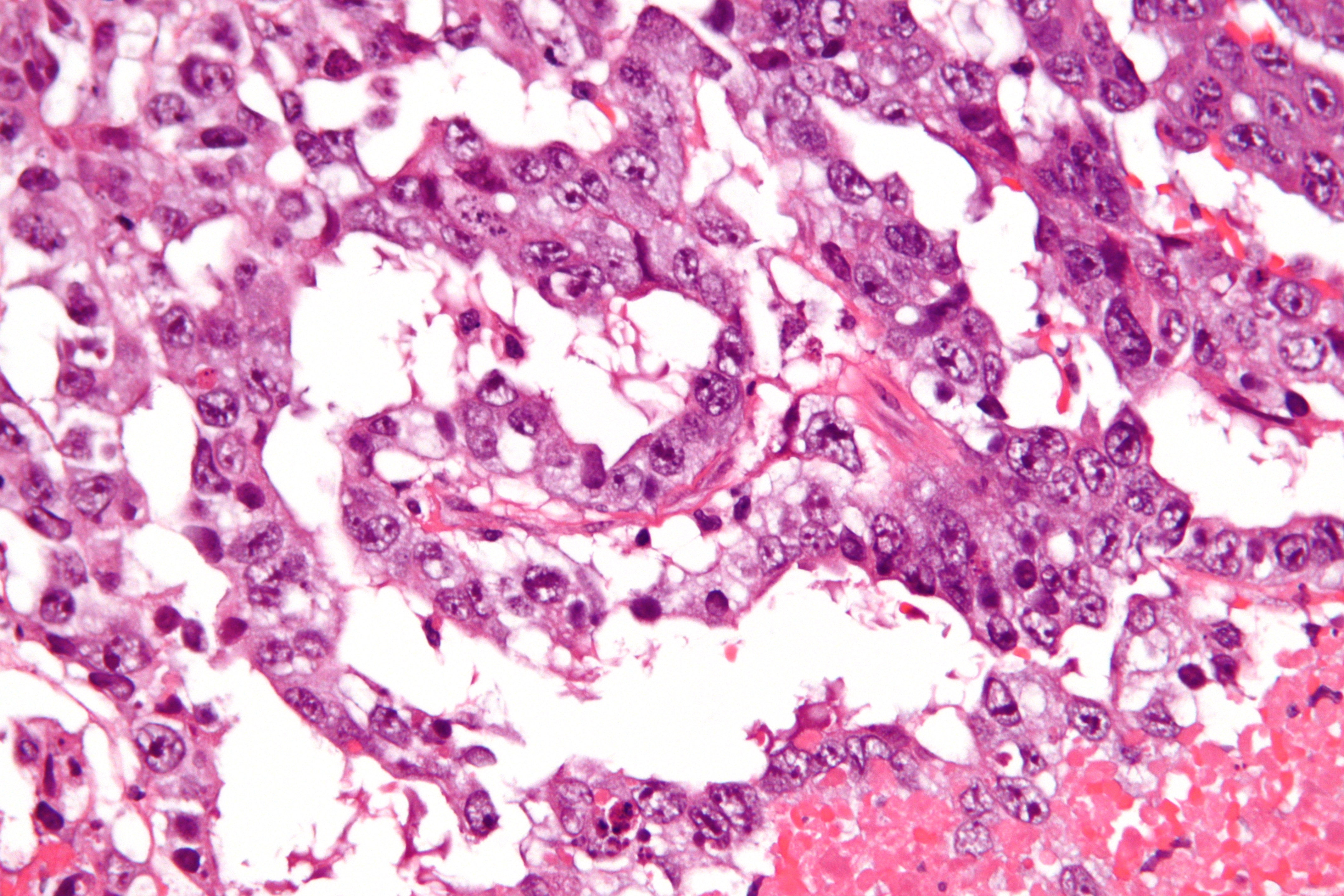
| Description | Very high magnification micrograph of a mixed germ cell tumour, also spelled mixed germ cell tumor. Testis (not apparent in images). H&E stain.
The intermediate magnification image show both embryonal carcinoma (right of image) and yolk sac tumour (left of image). The higher magnification images show only yolk sac tumour. Common mixed germ cell tumour combinations are: Teratoma and embryonal carcinoma and yolk sac tumour. Seminoma and embryonal carcinoma. Teratoma and embryonal carcinoma. Yolk sac tumour has ten different morphologic patterns: Reticular. Endodermal sinus-like. Microcystic. Papillary. Solid. Glandular. Alveolar Polyvesicular vitelline. Enteric. Hepatoid. Related images
Intermed. mag.
High mag.
|
| Source | Wikimedia Commons file page |
| Author | Nephron |
| Permission | See original Commons license details. |
Licensing[edit]
Creative Commons Attribution-ShareAlike 3.0 Unported (CC BY-SA 3.0)
This file is licensed under the Creative Commons Attribution-ShareAlike 3.0 license.
Official license: CC BY-SA 3.0
License page: CC BY-SA 3.0
Original attribution and file history: Wikimedia Commons
File history
Click on a date/time to view the file as it appeared at that time.
| Date/Time | Thumbnail | Dimensions | User | Comment | |
|---|---|---|---|---|---|
| current | 03:36, 5 June 2026 | | 4,272 × 2,848 (4.38 MB) | Maintenance script (talk | contribs) | == Summary == Importing file |
You cannot overwrite this file.
File usage
The following 2 pages use this file:
