File:Myxomatous aortic valve.jpg
Original file (2,048 × 1,536 pixels, file size: 342 KB, MIME type: image/jpeg)
Summary[edit]
| Summary | |
|---|---|
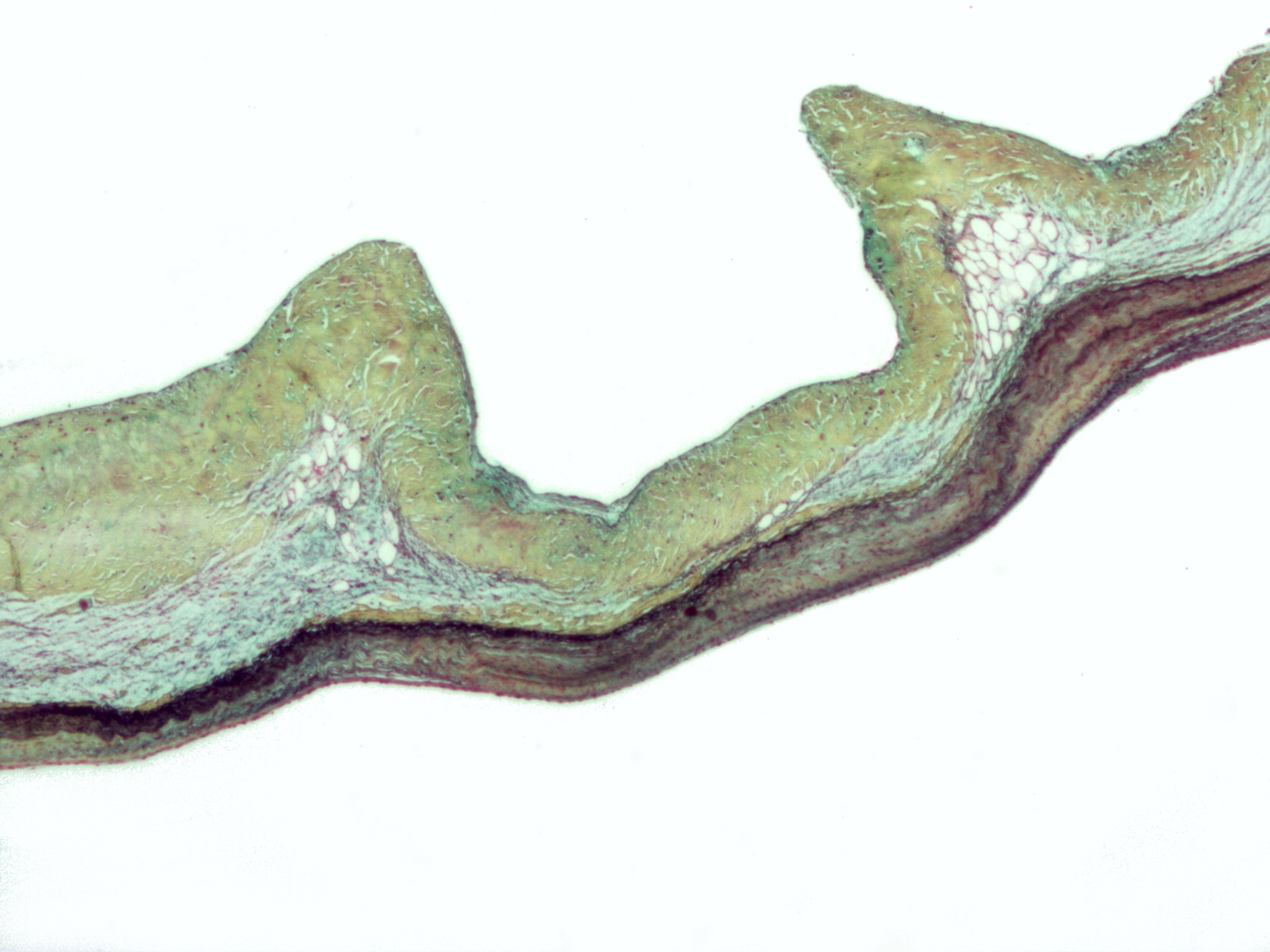
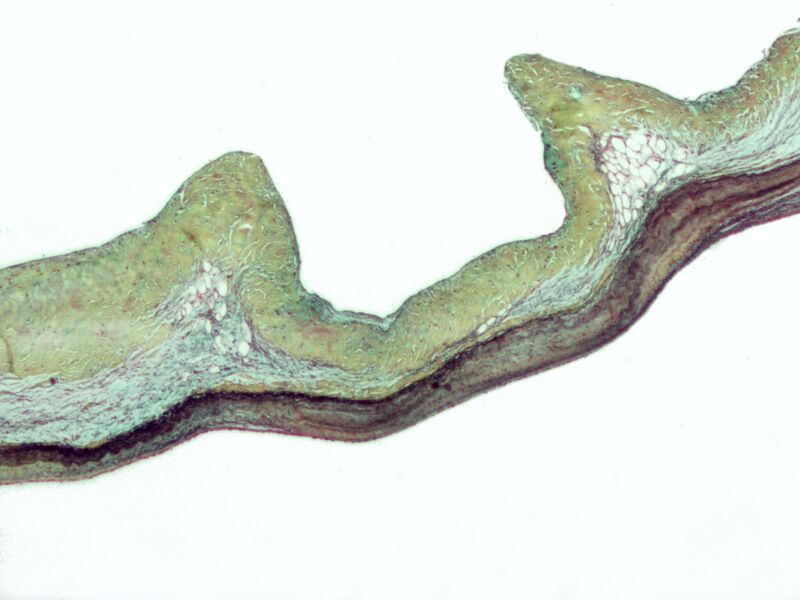
| Description | Micrograph of myxomatous degeneration of the aortic valve. Surgical specimen. Movat's stain (Black = nuclei, elastic fibres. Yellow = collagen, reticular fibers. Blue =
ground substance, mucin. Bright red = Fibrin. Red = muscle.) In myxomatous degeneration, the ventricularis layer (composed primarily of elastic tissue) is thinned and the spongiosa layer (composed of loose connective tissue) is thickened. On the image, the fibrosa layer (composed of collagen) is on the top, the thickened spongiosa layer below it and the ventricularis layer (made of elastic tissue) at the bottom. The ventricularis layer, as the name may suggest, is closest to the (left) ventricle. The fibrosa layer is closest to the sinus of valsalva. See also Marfan's syndrome - a condition, due to a defect in fibrillin (an essential component of elastic fibers), in which myxomatous degeneration is common. |
| Source | Wikimedia Commons file page |
| Author | Nephron |
| Permission | See original Commons license details. |
Licensing[edit]
Creative Commons Attribution-ShareAlike 3.0 Unported (CC BY-SA 3.0)
This file is licensed under the Creative Commons Attribution-ShareAlike 3.0 license.
Official license: CC BY-SA 3.0
License page: CC BY-SA 3.0
Original attribution and file history: Wikimedia Commons
File history
Click on a date/time to view the file as it appeared at that time.
| Date/Time | Thumbnail | Dimensions | User | Comment | |
|---|---|---|---|---|---|
| current | 03:18, 5 June 2026 | | 2,048 × 1,536 (342 KB) | Maintenance script (talk | contribs) | == Summary == Importing file |
You cannot overwrite this file.
File usage
The following file is a duplicate of this file (more details):
- File:Myxomatous aortic valve.jpg from Wikimedia Commons
The following 3 pages use this file:
