File:Spermatocytic seminoma high mag.jpg
Original file (4,272 × 2,848 pixels, file size: 3.75 MB, MIME type: image/jpeg)
Summary[edit]
| Summary | |
|---|---|
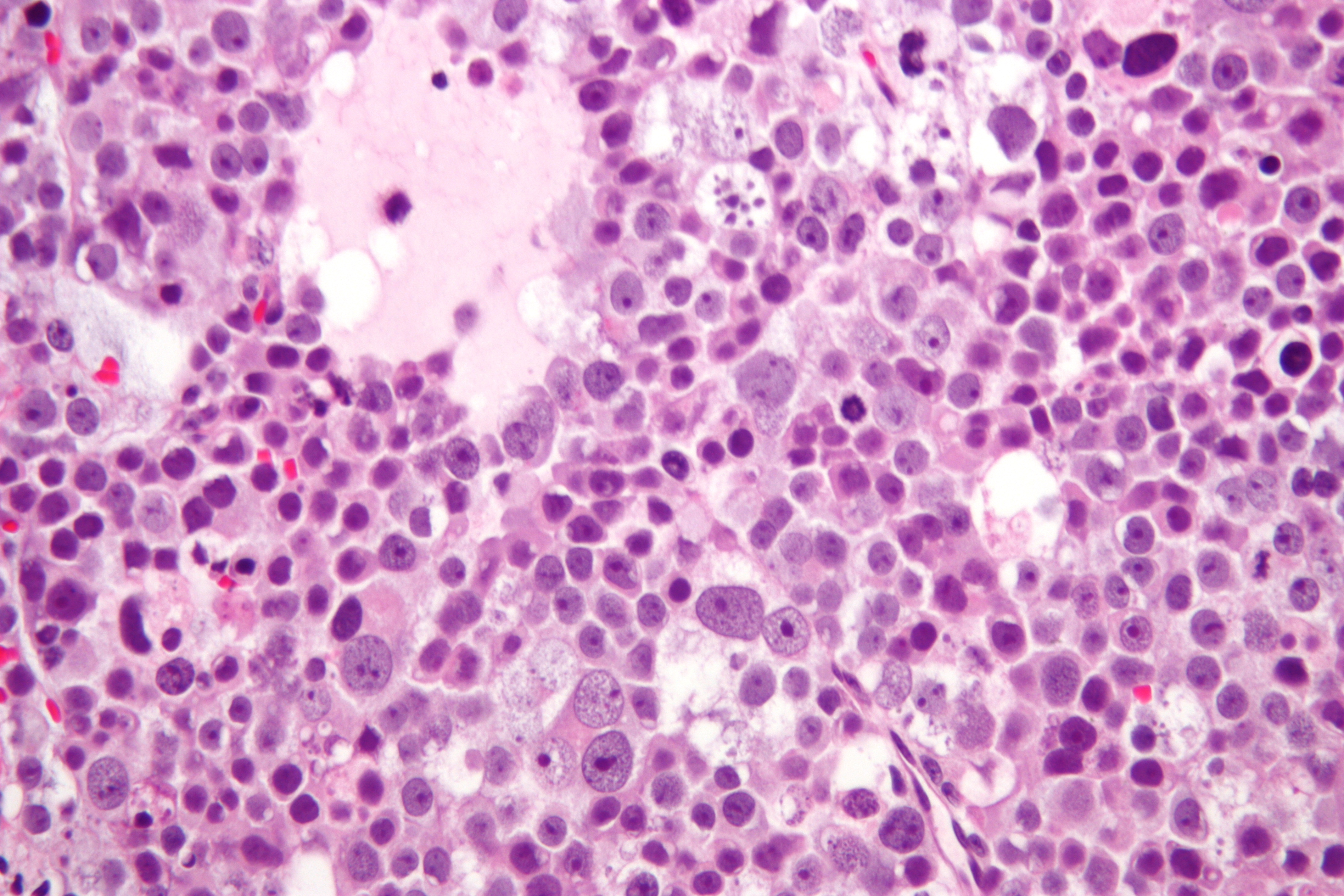
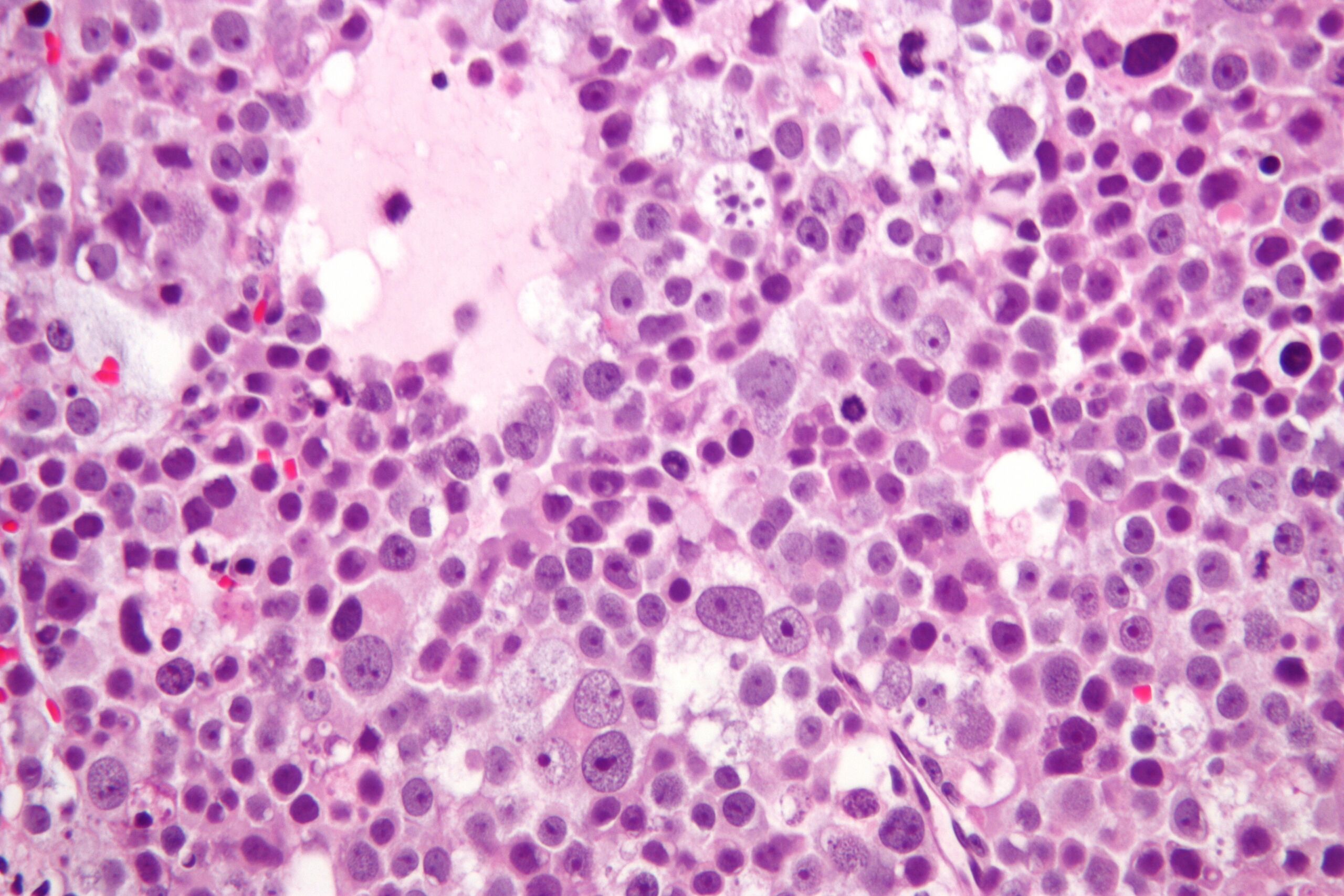
| Description | Micrograph of a spermatocytic seminoma. H&E stain.
Features of spermatocytic seminoma: Population of three cells. Small cells (6-8 µm) - with a large NC ratio. Look like secondary spermatocytes. Medium cells (15-18 µm) with prominent nucleoli. Filamentous chromatin (AKA spireme chromatin). Large cells (50-100 µm). Filamentous chromatin. Mucoid lakes. Intratubular spread (not seen on this image). Related images
|
| Source | Wikimedia Commons file page |
| Author | Nephron |
| Permission | See original Commons license details. |
Licensing[edit]
Creative Commons Attribution-ShareAlike 3.0 Unported (CC BY-SA 3.0)
This file is licensed under the Creative Commons Attribution-ShareAlike 3.0 license.
Official license: CC BY-SA 3.0
License page: CC BY-SA 3.0
Original attribution and file history: Wikimedia Commons
File history
Click on a date/time to view the file as it appeared at that time.
| Date/Time | Thumbnail | Dimensions | User | Comment | |
|---|---|---|---|---|---|
| current | 03:37, 5 June 2026 | | 4,272 × 2,848 (3.75 MB) | Maintenance script (talk | contribs) | == Summary == Importing file |
You cannot overwrite this file.
File usage
The following 2 pages use this file:
