File:Sucking chest wound mechanics 2.jpg
From WikiMD's WELLNESSPEDIA
No higher resolution available.
Sucking_chest_wound_mechanics_2.jpg (248 × 419 pixels, file size: 34 KB, MIME type: image/jpeg)
Summary[edit]
| Summary | |
|---|---|
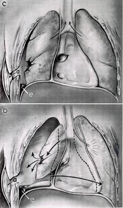
| Description | caption reads: FIGURE 2.—Continued. C. Packing of sucking wound (a), after which respiration becomes more normal. Hemothorax (e). D. Development of tension pneumothorax because air cannot escape from tear in lung (a), after wound is adequately packed. If it develops, it must be treated by closed (catheter) drainage of cavity. Hemothorax (e). |
| Source | Wikimedia Commons file page |
| Author | Lyman A. Brewer III, M.D., and Thomas H. Burford, M.D. |
| Permission | See original Commons license details. |
Licensing[edit]
Public Domain
This file is in the public domain and may be used without restriction.
Please see the linked source page for the original file history, attribution information, and licensing details.
Original attribution and file history: Wikimedia Commons
File history
Click on a date/time to view the file as it appeared at that time.
| Date/Time | Thumbnail | Dimensions | User | Comment | |
|---|---|---|---|---|---|
| current | 22:30, 8 June 2026 | | 248 × 419 (34 KB) | Maintenance script (talk | contribs) | == Summary == Importing file |
You cannot overwrite this file.
File usage
The following file is a duplicate of this file (more details):
- File:Sucking chest wound mechanics 2.jpg from Wikimedia Commons
The following page uses this file:
