File:The science and art of midwifery (1891) (14579471209).jpg
Original file (744 × 1,912 pixels, file size: 216 KB, MIME type: image/jpeg)
Summary[edit]
| Summary | |
|---|---|
| Description | Identifier: scienceamidw00lusk (find matches)
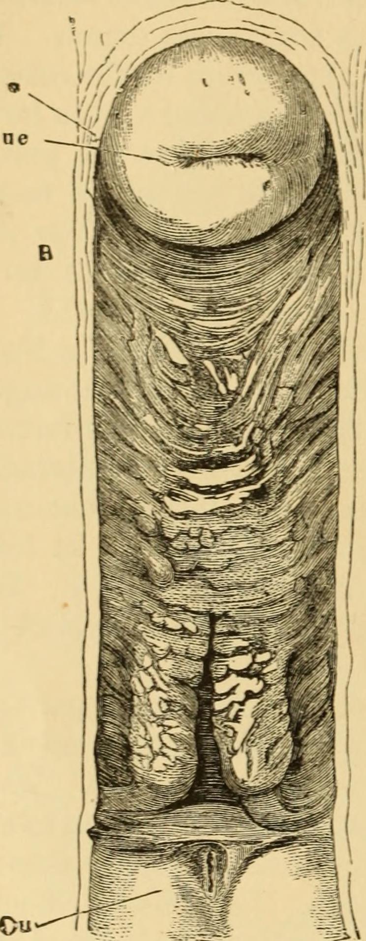
Title: The science and art of midwifery Year: 1891 (1890s) Authors: Lusk, William Thompson, 1838-1897 Subjects: Obstetrics Publisher: New York, D. Appleton and Co. Contributing Library: Yale University, Cushing/Whitney Medical Library Digitizing Sponsor: Open Knowledge Commons and Yale University, Cushing/Whitney Medical Library View Book Page: Book Viewer About This Book: Catalog Entry View All Images: All Images From Book Click here to view book online to see this illustration in context in a browseable online version of this book. Text Appearing Before Image: velopmentas to communicate to the finger a dis-tinctly granular sensation. Though the secreting glands of thevagina f are few in number, it is covered,even in periods of repose, with a thinlayer of acid mucus. Under sexual ex-citement, and during menstruation orpregnancy, the amount of this secretionis largely increased. The hypogastric, the uterine, thevesical, and the pudendal arteries all send branches to the vagina.The pulsations of the uterine artery may sometimes be felt throughthe upper part of the vaginal walls. During pregnancy these pulsa-tions are always so distinctly marked as to constitute a good inferen-tial sign of that condition. The veins form a close plexus around the vagina. Like all thepelvic veins, they are without valves, and are therefore peculiarly sub- * Henle, Handbuch dcr Eingeweidelehre des Menscben, Braunschweig, 1S66, p.450. f Tbe occasional presence of glands in the vagina has been demonstrated by Preus-chen. Vide Yirchows Archiv, 1877, vol. lxx, p. 111. Text Appearing After Image: Fig. 8.—The vacrina (exposed in itsentire length by the removal ofthe posterior wall). Ou. orifici-um urethra?; Oue, orificium utc-rinum-externurn; B. section ofwall at the fornix vaginas. (Hen-le.) FEMALE ORGANS OF GENERATION, 11 Note About Images Please note that these images are extracted from scanned page images that may have been digitally enhanced for readability - coloration and appearance of these illustrations may not perfectly resemble the original work. |
| Source | Wikimedia Commons file page |
| Author | Internet Archive Book Images |
| Permission | See original Commons license details. |
Licensing[edit]
License: No restrictions
License page: No restrictions
Original attribution and file history: Wikimedia Commons
File history
Click on a date/time to view the file as it appeared at that time.
| Date/Time | Thumbnail | Dimensions | User | Comment | |
|---|---|---|---|---|---|
| current | 22:31, 8 June 2026 | 744 × 1,912 (216 KB) | Maintenance script (talk | contribs) | == Summary == Importing file |
You cannot overwrite this file.
File usage
The following page uses this file:

_(14579471209).jpg&action=edit§ion=1)
_(14579471209).jpg)
_(14579471209).jpg&action=edit§ion=2)
_(14579471209).jpg&oldid=6612708)