File:Wernicke's area animation.gif
From WikiMD's WELLNESSPEDIA
No higher resolution available.
Wernicke's_area_animation.gif (300 × 300 pixels, file size: 1.99 MB, MIME type: image/gif, looped, 72 frames, 10 s)
Summary[edit]
| Summary | |
|---|---|
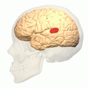
| Description | Wernicke's area (shown in red).Colored region is posterior section of the superior temporal gyrus (pSTG) of the left cerebral hemisphere. Though this region is generally treated as Wernicke's area, there are many researches and discussions about its exact size and anatomical boundaries.
Wise RJ, Scott SK, Blank SC, Mummery CJ, Murphy K, Warburton EA. "Separate neural subsystems within 'Wernicke's area'. " Brain: 2001, 124(Pt 1);83-95 PMID 11133789 Tomoo Inubushi, Kuniyoshi Sakai (2014) "Language center" Brain Science Dictionary http://bsd.neuroinf.jp/wiki/%E8%A8%80%E8%AA%9E%E4%B8%AD%E6%9E%A2 (in Japanese) |
| Source | Wikimedia Commons file page |
| Author | Polygon data were generated by Database Center for Life Science(DBCLS)[2]. |
| Permission | See original Commons license details. |
Licensing[edit]
License: CC BY-SA 2.1 jp
License page: CC BY-SA 2.1 jp
Original attribution and file history: Wikimedia Commons
File history
Click on a date/time to view the file as it appeared at that time.
| Date/Time | Thumbnail | Dimensions | User | Comment | |
|---|---|---|---|---|---|
| current | 12:49, 29 May 2026 | | 300 × 300 (1.99 MB) | Maintenance script (talk | contribs) | == Summary == Importing file |
You cannot overwrite this file.
File usage
The following 2 pages use this file:
