File:Zona pellucida.jpg
From WikiMD's medical encyclopedia
Size of this preview: 600 × 600 pixels. Other resolutions: 240 × 240 pixels | 480 × 480 pixels | 768 × 768 pixels | 1,024 × 1,024 pixels | 2,048 × 2,048 pixels.
Original file (2,048 × 2,048 pixels, file size: 1.51 MB, MIME type: image/jpeg)
Summary
| Description |
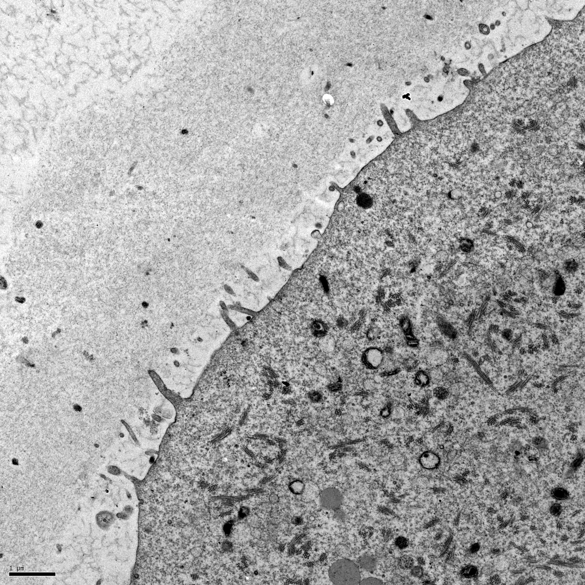
Türkçe: Zonna pellucida (human).
Surface of a mouse oocyte This transmission electron micrograph shows the cortex of a mouse oocyte that was undergoing fertilization. The sample was fixed using glutaraldehyde and osmium tetroxide, embedded in plastic, sectioned, and stained with uranyl acetate and lead citrate. Microvilli are present on the oocyte's surface and the zona pellucida is visible surrounding the oocyte. The image was taken with a Phillips 500 transmission electron microscope. The scale bar is 1. |
| Date | |
| Source | http://www.cellimagelibrary.org/images/12618 |
| Author | Prue Talbot |
Licensing
| This file is made available under the Creative Commons CC0 1.0 Universal Public Domain Dedication. | |
| The person who associated a work with this deed has dedicated the work to the public domain by waiving all of their rights to the work worldwide under copyright law, including all related and neighboring rights, to the extent allowed by law. You can copy, modify, distribute and perform the work, even for commercial purposes, all without asking permission.
|
File history
Click on a date/time to view the file as it appeared at that time.
| Date/Time | Thumbnail | Dimensions | User | Comment | |
|---|---|---|---|---|---|
| current | 21:45, 26 March 2014 | | 2,048 × 2,048 (1.51 MB) | Ayrıntılı Bilgi | User created page with UploadWizard |
File usage
The following page uses this file: