Gram stain
Gram stain is a method of staining used in microbiology to differentiate bacterial species into two large groups (Gram-positive and Gram-negative). The name comes from the Danish bacteriologist Hans Christian Gram, who developed the technique.
History[edit]
Hans Christian Gram developed the Gram stain in 1884 to differentiate bacterial cells in lung tissue samples. He noticed that certain stains were retained by some cells but not others, leading to the classification of bacteria into Gram-positive and Gram-negative.
Procedure[edit]
The Gram stain procedure involves four steps:
- Application of a primary stain (Crystal Violet)
- Application of a mordant (Gram's Iodine)
- Decolorization with alcohol or acetone
- Counterstaining with a secondary stain (Safranin)
Interpretation[edit]
Gram-positive bacteria retain the crystal violet dye, and thus they are stained violet, while the Gram-negative bacteria do not retain this stain and are thus stained red from the safranin counterstain.
Applications[edit]
The Gram stain is almost always the first step in the preliminary identification of a bacterial organism. It helps in the classification and differentiation of microorganisms. It is also used in the initial steps of diagnosing an infection.
Limitations[edit]
While Gram staining is a valuable diagnostic tool in both clinical and research settings, not all bacteria can be definitively classified by this technique, these are known as Gram-variable and Gram-indeterminate.
See also[edit]
References[edit]

This WikiMD article can only be edited by registered and verified editors. You can log in or register.
Gram_stain[edit]
-
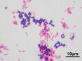
 Gram positive coccus and gram negative rod
 -
 Gram stain 01
 -
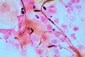 Candida Gram stain
 -
 Gram Staining Bacteria
 -
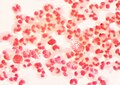
 Gram-positive bacteria and pus cells
 -
 Neisseria gonorrhoeae and pus cells Gram stain


Medical Disclaimer: WikiMD is for informational purposes only and is not a substitute for professional medical advice. Content may be inaccurate or outdated and should not be used for diagnosis or treatment. Always consult your healthcare provider for medical decisions. Verify information with trusted sources such as CDC.gov and NIH.gov. By using this site, you agree that WikiMD is not liable for any outcomes related to its content. See full disclaimer.
Credits:Most images are courtesy of Wikimedia commons, and templates, categories Wikipedia, licensed under CC BY SA or similar.
Translate page: - East Asian
中文,
日本,
한국어,
South Asian
हिन्दी,
தமிழ்,
తెలుగు,
Urdu,
ಕನ್ನಡ,
Southeast Asian
Indonesian,
Vietnamese,
Thai,
မြန်မာဘာသာ,
বাংলা
European
español,
Deutsch,
français,
Greek,
português do Brasil,
polski,
română,
русский,
Nederlands,
norsk,
svenska,
suomi,
Italian
Middle Eastern & African
عربى,
Turkish,
Persian,
Hebrew,
Afrikaans,
isiZulu,
Kiswahili,
Other
Bulgarian,
Hungarian,
Czech,
Swedish,
മലയാളം,
मराठी,
ਪੰਜਾਬੀ,
ગુજરાતી,
Portuguese,
Ukrainian
