Ciliary ganglion
The ciliary ganglion is a type of parasympathetic ganglion located in the orbit behind the eye and lateral to the optic nerve. It is approximately 2mm in size and receives input from the oculomotor nerve and provides output to the ciliary muscles and constrictor pupillae.
Anatomy[edit]
The ciliary ganglion is a small, flattened structure that is suspended by two roots: the sensory root from the nasociliary nerve and the motor root from the inferior division of the oculomotor nerve. The ganglion is located in the posterior part of the orbit. It is situated between the optic nerve and the lateral rectus muscle, closer to the latter.
Function[edit]
The ciliary ganglion serves as a relay station for parasympathetic fibers traveling to the eye. It receives preganglionic parasympathetic fibers from the Edinger-Westphal nucleus via the oculomotor nerve. These fibers synapse in the ganglion with postganglionic fibers that innervate the ciliary muscles, which control the shape of the lens, and the constrictor pupillae, which control the size of the pupil.
Clinical significance[edit]
Damage to the ciliary ganglion can result in a condition known as Adie's pupil, characterized by a dilated pupil that responds slowly to light but reacts normally to accommodation. Other potential conditions include ophthalmoplegia, ptosis, and anisocoria.
See also[edit]
- Parasympathetic nervous system
- Sympathetic nervous system
- Autonomic ganglion
- Oculomotor nerve
- Edinger-Westphal nucleus
References[edit]

This article about a neurology is a stub. You can help WikiMD by expanding the page. |
This ophthalmology article is a stub. You can help WikiMD by expanding the page. |
Ciliary ganglion[edit]
-
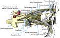 Diagram showing the nerves of the eye, including the ciliary ganglion.
 -
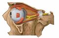 Lateral view of the orbit nerves, highlighting the ciliary ganglion.
 -
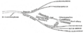
 Illustration of Horner's Syndrome and autonomic innervation of the eye, including the ciliary ganglion.
 -
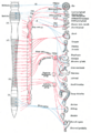
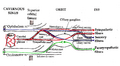 Pathways of the ciliary ganglion.
 -
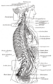
 Anatomical illustration from Gray's Anatomy showing the ciliary ganglion.
 -
 Gray's Anatomy illustration of the ciliary ganglion and associated structures.
 -
 Detailed view of the ciliary ganglion from Gray's Anatomy.


Medical Disclaimer: WikiMD is for informational purposes only and is not a substitute for professional medical advice. Content may be inaccurate or outdated and should not be used for diagnosis or treatment. Always consult your healthcare provider for medical decisions. Verify information with trusted sources such as CDC.gov and NIH.gov. By using this site, you agree that WikiMD is not liable for any outcomes related to its content. See full disclaimer.
Credits:Most images are courtesy of Wikimedia commons, and templates, categories Wikipedia, licensed under CC BY SA or similar.
Translate page: - East Asian
中文,
日本,
한국어,
South Asian
हिन्दी,
தமிழ்,
తెలుగు,
Urdu,
ಕನ್ನಡ,
Southeast Asian
Indonesian,
Vietnamese,
Thai,
မြန်မာဘာသာ,
বাংলা
European
español,
Deutsch,
français,
Greek,
português do Brasil,
polski,
română,
русский,
Nederlands,
norsk,
svenska,
suomi,
Italian
Middle Eastern & African
عربى,
Turkish,
Persian,
Hebrew,
Afrikaans,
isiZulu,
Kiswahili,
Other
Bulgarian,
Hungarian,
Czech,
Swedish,
മലയാളം,
मराठी,
ਪੰਜਾਬੀ,
ગુજરાતી,
Portuguese,
Ukrainian
