File:Crystal structure of human CD9.pdf
From WikiMD's medical encyclopedia
No higher resolution available.
Crystal_structure_of_human_CD9.pdf (750 × 600 pixels, file size: 1 MB, MIME type: application/pdf)
Summary
| Description |
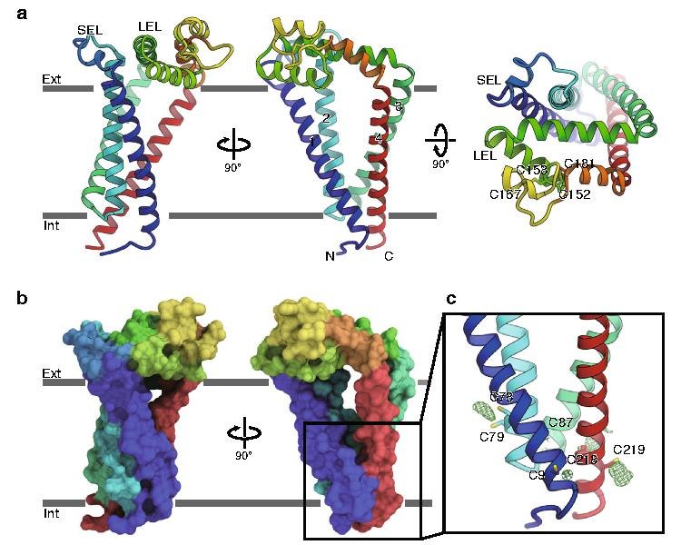
English: a Overall structure of human CD9, viewed from the membrane plane (left and middle) and from the extracellular side (right). The transmembrane helices and the two extracellular loops (SEL and LEL) are labeled. Cys152-Cys181 and Cys153-Cys167 form disulfide bonds. b Surface representation of CD9, colored according to a. c Palmitoylation of the cytoplasmic cysteine residues. Green meshes show Fo−Fc densities contoured at 2.5 σ, indicating the palmitoylation of the cysteine residues on the cytoplasmic end of the four transmembrane helices.[1] |
| Date | |
| Source | https://doi.org/10.1038/s41467-020-15459-7 |
| Author | Rie Umeda, Yuhkoh Satouh, Mizuki Takemoto, Yoshiko Nakada-Nakura, Kehong Liu, Takeshi Yokoyama, Mikako Shirouzu, So Iwata, Norimichi Nomura, Ken Sato, Masahito Ikawa, Tomohiro Nishizawa & Osamu Nureki |
Licensing
This file is licensed under the Creative Commons Attribution 4.0 International license.
- You are free:
- to share – to copy, distribute and transmit the work
- to remix – to adapt the work
- Under the following conditions:
- attribution – You must give appropriate credit, provide a link to the license, and indicate if changes were made. You may do so in any reasonable manner, but not in any way that suggests the licensor endorses you or your use.
File history
Click on a date/time to view the file as it appeared at that time.
| Date/Time | Thumbnail | Dimensions | User | Comment | |
|---|---|---|---|---|---|
| current | 22:47, 4 April 2020 |  | 750 × 600 (1 MB) | Rob Hurt | Uploaded a work by Rie Umeda, Yuhkoh Satouh, Mizuki Takemoto, Yoshiko Nakada-Nakura, Kehong Liu, Takeshi Yokoyama, Mikako Shirouzu, So Iwata, Norimichi Nomura, Ken Sato, Masahito Ikawa, Tomohiro Nishizawa & Osamu Nureki from https://doi.org/10.1038/s41467-020-15459-7 with UploadWizard |
File usage
The following page uses this file: