File:Nervous and mental diseases (1911) (14775178011).jpg
Original file (2,180 × 1,552 pixels, file size: 498 KB, MIME type: image/jpeg)
Summary[edit]
| Summary | |
|---|---|
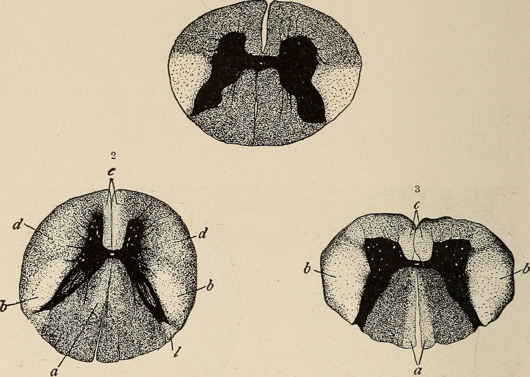
| Description | Identifier: nervousmentald00chur (find matches)
Title: Nervous and mental diseases Year: 1911 (1910s) Authors: Church, Archibald, b. 1861 Peterson, Frederick, 1859-1938, joint author Subjects: Nervous system Publisher: Philadelphia and London, W. B. Saunders company Contributing Library: The Library of Congress Digitizing Sponsor: The Library of Congress View Book Page: Book Viewer About This Book: Catalog Entry View All Images: All Images From Book Click here to view book online to see this illustration in context in a browseable online version of this book. Text Appearing Before Image: emotor and trophic portions of the central apparatus, and constitutes aprimordial shortcoming bv which these parts reach an earlv death. 26 402 DISEASES OF THE CORD PROPER. Morbid Anatomy.—The lesions of progressive spinal muscularatrophy embrace in rare cases the entire motor field of the nervousapparatus from cerebral cortex to muscular nerve-endings, and includethe muscles themselves. Both upper and lower motor neurons in theirentirety are destroyed by a degenerative process. Following the patholog-ical rule that a neuron degenerating from toxic cause or involution firstshows changes in its peripheral portion, the upper motor segment maypresent alteration only in the pyramidal fibers of the cord. This mayreach the medulla, and, as a rule, does not extend into the peduncles,capsule, and cortex, though it may do so. In the lower neuron the de-generation is probably at first peripheral, but in all cases that reach amarked development the cells of the anterior gray are found degener- Text Appearing After Image: Fig. 155.—Cord-sections in a case of amyotrophic lateral sclerosis. 1, Lumbar region; 2, dorsal region; 3, cervical region (Marie). ated. Attending this we have muscular atrophy, with fibroid and fattychanges and degeneration in the motor fibers of the nerve-trunks, limitedsharply by the anatomical relations of the diseased cord-elements. In the cord the gray substance of the anterior horns shows atrophy.The ganglion-cells, many of which usually have disappeared, are wastedand degenerated, and there is a general shrinking of all the nervous ele-ments of the horn. The white substance of both the direct and crossedpyramidal tracts shows sclerotic degeneration. This process is notstrictly confined to them, but usually involves the anterolateral tracts toa lesser degree, and may invade the lateral limiting layer. This is espe-cially the case in the upper dorsal and cervical regions. The columnsof Groll sometimes show slight changes, apparently due to the shrinkingof the myelin, and not Note About Images Please note that these images are extracted from scanned page images that may have been digitally enhanced for readability - coloration and appearance of these illustrations may not perfectly resemble the original work. |
| Source | Wikimedia Commons file page |
| Author | Internet Archive Book Images |
| Permission | See original Commons license details. |
Licensing[edit]
License: No restrictions
License page: No restrictions
Original attribution and file history: Wikimedia Commons
File history
Click on a date/time to view the file as it appeared at that time.
| Date/Time | Thumbnail | Dimensions | User | Comment | |
|---|---|---|---|---|---|
| current | 22:35, 8 June 2026 | | 2,180 × 1,552 (498 KB) | Maintenance script (talk | contribs) | == Summary == Importing file |
You cannot overwrite this file.
File usage
The following file is a duplicate of this file (more details):
- File:Nervous and mental diseases (1911) (14775178011).jpg from Wikimedia Commons
The following page uses this file:

_(14775178011).jpg&action=edit§ion=1)
_(14775178011).jpg)
_(14775178011).jpg&action=edit§ion=2)
_(14775178011).jpg)
_(14775178011).jpg&oldid=6610391)