File:Schematic diagram of the human eye en.svg
From WikiMD's medical encyclopedia

Size of this PNG preview of this SVG file: 416 × 423 pixels. Other resolutions: 236 × 240 pixels | 472 × 480 pixels | 590 × 600 pixels | 755 × 768 pixels | 1,007 × 1,024 pixels | 2,014 × 2,048 pixels.
Original file (SVG file, nominally 416 × 423 pixels, file size: 319 KB)
Summary
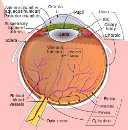
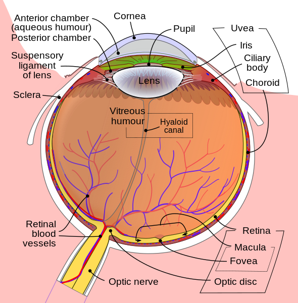
| Description | Diagram of the human eye in English. It shows the lower part of the right eye after a central and horizontal section. |
| Date | |
| Source | Schematic_diagram_of_the_human_eye_with_English_annotations.svg |
| Author | Rhcastilhos. And Jmarchn. |
| Other versions |
[]
By languages
For translate
Anterior segment |
| SVG development |


Licensing
I, the copyright holder of this work, hereby publish it under the following license:
This file is licensed under the Creative Commons Attribution-Share Alike 3.0 Unported license.
- You are free:
- to share – to copy, distribute and transmit the work
- to remix – to adapt the work
- Under the following conditions:
- attribution – You must give appropriate credit, provide a link to the license, and indicate if changes were made. You may do so in any reasonable manner, but not in any way that suggests the licensor endorses you or your use.
- share alike – If you remix, transform, or build upon the material, you must distribute your contributions under the same or compatible license as the original.
File history
Click on a date/time to view the file as it appeared at that time.
| Date/Time | Thumbnail | Dimensions | User | Comment | |
|---|---|---|---|---|---|
| current | 15:35, 6 October 2024 | | 416 × 423 (319 KB) | TiberiuFr25 | File uploaded using svgtranslate tool (https://svgtranslate.toolforge.org/). Added translation for ro. |
File usage
The following 26 pages use this file:
- Anterior chamber of eyeball
- Globe (human eye)
- Human eye
- Hyaloid artery
- Hyaloid canal
- Intraocular hemorrhage
- Iris (anatomy)
- Macular telangiectasia
- Ocular immune system
- Optic disc
- Periocular injection
- Persistent tunica vasculosa lentis
- Pigment dispersion syndrome
- Posterior segment of eyeball
- Posterior vitreous detachment
- Pupil
- Stimulus modality
- Subconjunctival injection
- Suprachoroidal drug delivery
- Supratarsal injection
- Synchysis scintillans
- The Organ of Sight
- Vitreomacular adhesion
- Vitreous body
- Vitreous chamber
- Vitreous membrane