Iris (anatomy)
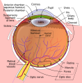
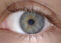
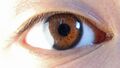
The iris is a thin, circular structure in the eye, responsible for controlling the diameter and size of the pupil and thus the amount of light reaching the retina. The color of the iris gives the eye its color.
Structure[edit]
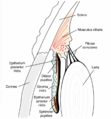
The iris consists of two main layers: the front pigmented fibrovascular known as a stroma and, beneath the stroma, pigmented epithelial cells.
Stroma[edit]
The stroma connects to a sphincter muscle (sphincter pupillae), which contracts the pupil in a circular motion, and a set of dilator muscles (dilator pupillae) which pull the iris radially to enlarge the pupil, pulling it in folds.
Pigmented epithelial cells[edit]
The pigmented epithelial cells, lying behind the stroma, are a double layer of columnar cells that secrete and absorb aqueous humor.
Function[edit]
The main function of the iris is to control the amount of light getting into the eye. This is done by adjusting the size of the pupil, which is the hole in the middle of the iris through which light enters the eye.
Clinical significance[edit]
Diseases such as aniridia, coloboma, and iritis can affect the iris.
See also[edit]
References[edit]
| Anatomy of the globe of the human eye | ||||||||||||||||||
|---|---|---|---|---|---|---|---|---|---|---|---|---|---|---|---|---|---|---|
|
| Human eye anatomy and physiology | ||||||||||
|---|---|---|---|---|---|---|---|---|---|---|
|
This medical article is a stub. You can help WikiMD by expanding the page. |
-
 Iris (anatomy)
 -
 Iris (anatomy)
 -
 Iris (anatomy)
 -
Iris (anatomy)
-
Iris (anatomy)
-
Iris (anatomy)
-
 Iris (anatomy)
 -
 Iris (anatomy)
 -
 Iris (anatomy)
 -
Iris (anatomy)
-
 Iris (anatomy)
 -
 Iris (anatomy)


.png)


")
Medical Disclaimer: WikiMD is for informational purposes only and is not a substitute for professional medical advice. Content may be inaccurate or outdated and should not be used for diagnosis or treatment. Always consult your healthcare provider for medical decisions. Verify information with trusted sources such as CDC.gov and NIH.gov. By using this site, you agree that WikiMD is not liable for any outcomes related to its content. See full disclaimer.
Credits:Most images are courtesy of Wikimedia commons, and templates, categories Wikipedia, licensed under CC BY SA or similar.
Translate page: - East Asian
中文,
日本,
한국어,
South Asian
हिन्दी,
தமிழ்,
తెలుగు,
Urdu,
ಕನ್ನಡ,
Southeast Asian
Indonesian,
Vietnamese,
Thai,
မြန်မာဘာသာ,
বাংলা
European
español,
Deutsch,
français,
Greek,
português do Brasil,
polski,
română,
русский,
Nederlands,
norsk,
svenska,
suomi,
Italian
Middle Eastern & African
عربى,
Turkish,
Persian,
Hebrew,
Afrikaans,
isiZulu,
Kiswahili,
Other
Bulgarian,
Hungarian,
Czech,
Swedish,
മലയാളം,
मराठी,
ਪੰਜਾਬੀ,
ગુજરાતી,
Portuguese,
Ukrainian
