Parafovea
Parafovea is a region of the retina that surrounds the fovea. It is a part of the macula and is responsible for the second highest resolution of vision in the eye. The parafovea is located approximately 1.25 to 2.75 mm from the center of the fovea.
Anatomy[edit]
The parafovea is a circular area that surrounds the fovea and is part of the macula. It is approximately 2.5 mm in diameter. The parafovea is made up of four layers: the ganglion cell layer, the inner plexiform layer, the inner nuclear layer, and the outer plexiform layer. The density of photoreceptor cells in the parafovea is less than that in the fovea, but greater than that in the rest of the retina.
Function[edit]
The parafovea is responsible for high-resolution vision, second only to the fovea. It is particularly important for color vision and for seeing fine detail. The parafovea is also involved in peripheral vision, which is the ability to see objects outside the direct line of sight.
Clinical significance[edit]
Diseases that affect the parafovea can lead to vision loss. These include macular degeneration, diabetic retinopathy, and retinal detachment. Early detection and treatment of these conditions can help to preserve vision.
See also[edit]
| Anatomy of the globe of the human eye | ||||||||||||||||||
|---|---|---|---|---|---|---|---|---|---|---|---|---|---|---|---|---|---|---|
|
| Vision | ||||||||||
|---|---|---|---|---|---|---|---|---|---|---|
This Vision related articles is a stub.
|
| Human eye anatomy and physiology | ||||||||||
|---|---|---|---|---|---|---|---|---|---|---|
|

This WikiMD article can only be edited by registered and verified editors. You can log in or register.
-
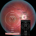 Diagram of the macula
 -
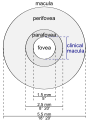 Illustration of the macula lutea
 -
 Optical coherence tomography of the retina
 -
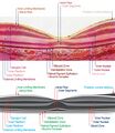
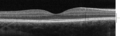 Spectral domain OCT cross-section of the macula
 -
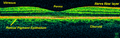
 Histological image of the macula
 -
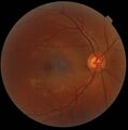 Retinography image


Medical Disclaimer: WikiMD is for informational purposes only and is not a substitute for professional medical advice. Content may be inaccurate or outdated and should not be used for diagnosis or treatment. Always consult your healthcare provider for medical decisions. Verify information with trusted sources such as CDC.gov and NIH.gov. By using this site, you agree that WikiMD is not liable for any outcomes related to its content. See full disclaimer.
Credits:Most images are courtesy of Wikimedia commons, and templates, categories Wikipedia, licensed under CC BY SA or similar.
Translate page: - East Asian
中文,
日本,
한국어,
South Asian
हिन्दी,
தமிழ்,
తెలుగు,
Urdu,
ಕನ್ನಡ,
Southeast Asian
Indonesian,
Vietnamese,
Thai,
မြန်မာဘာသာ,
বাংলা
European
español,
Deutsch,
français,
Greek,
português do Brasil,
polski,
română,
русский,
Nederlands,
norsk,
svenska,
suomi,
Italian
Middle Eastern & African
عربى,
Turkish,
Persian,
Hebrew,
Afrikaans,
isiZulu,
Kiswahili,
Other
Bulgarian,
Hungarian,
Czech,
Swedish,
മലയാളം,
मराठी,
ਪੰਜਾਬੀ,
ગુજરાતી,
Portuguese,
Ukrainian
