Gamma camera
A gamma camera (also known as a scintillation camera) is a device used in nuclear medicine imaging to capture the radiation from a patient's body in order to produce images for diagnostic purposes. The gamma camera is a key tool in the field of medical imaging and is used in a variety of medical procedures, including single photon emission computed tomography (SPECT) and positron emission tomography (PET).
History[edit]
The gamma camera was first developed in the 1950s by Hal Anger, an American electrical engineer and biophysicist. Anger's original design has been refined and improved over the years, but the basic principles of operation remain the same.
Operation[edit]
The gamma camera operates by detecting gamma rays, which are high-energy photons emitted by radioactive substances. The patient is injected with a small amount of a radioactive tracer, which travels through the body and emits gamma rays. The gamma camera detects these rays and uses them to create an image of the area of the body being examined.
The main components of a gamma camera are the collimator, the scintillator, and the photomultiplier tubes. The collimator is a lead shield with many small holes, which allows only gamma rays traveling in certain directions to reach the scintillator. The scintillator is a crystal that emits light when struck by gamma rays. The photomultiplier tubes convert this light into an electrical signal, which is then processed to create an image.
Applications[edit]
Gamma cameras are used in a wide range of medical procedures. They are most commonly used in nuclear medicine imaging, where they can provide detailed images of the body's organs and tissues. This can be used to diagnose a variety of conditions, including cancer, heart disease, and thyroid disorders.
In addition to their use in medical imaging, gamma cameras are also used in industrial radiography and radiation therapy.
See Also[edit]
- Nuclear medicine
- Medical imaging
- Single photon emission computed tomography
- Positron emission tomography
- Radioactive tracer
- Collimator
- Scintillator
- Photomultiplier tube
This medical article is a stub. You can help WikiMD by expanding the page. |
This radiology related article is a stub. You can help WikiMD by expanding it.
Gamma camera[edit]
-
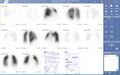 Lung scintigraphy
 -
Coded aperture mask for gamma camera
-
Gamma camera
-
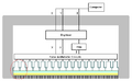 Gamma camera cross section
 -
Gamma camera cross section detail
-
Collimated and penetration


Medical Disclaimer: WikiMD is for informational purposes only and is not a substitute for professional medical advice. Content may be inaccurate or outdated and should not be used for diagnosis or treatment. Always consult your healthcare provider for medical decisions. Verify information with trusted sources such as CDC.gov and NIH.gov. By using this site, you agree that WikiMD is not liable for any outcomes related to its content. See full disclaimer.
Credits:Most images are courtesy of Wikimedia commons, and templates, categories Wikipedia, licensed under CC BY SA or similar.
Translate page: - East Asian
中文,
日本,
한국어,
South Asian
हिन्दी,
தமிழ்,
తెలుగు,
Urdu,
ಕನ್ನಡ,
Southeast Asian
Indonesian,
Vietnamese,
Thai,
မြန်မာဘာသာ,
বাংলা
European
español,
Deutsch,
français,
Greek,
português do Brasil,
polski,
română,
русский,
Nederlands,
norsk,
svenska,
suomi,
Italian
Middle Eastern & African
عربى,
Turkish,
Persian,
Hebrew,
Afrikaans,
isiZulu,
Kiswahili,
Other
Bulgarian,
Hungarian,
Czech,
Swedish,
മലയാളം,
मराठी,
ਪੰਜਾਬੀ,
ગુજરાતી,
Portuguese,
Ukrainian

.jpg)