Magnetic resonance imaging
| Pronunciation | |
|---|---|
| Other names | |
| Medical specialty | |
| Uses | |
| Complications | |
| Approach | |
| Types | |
| Recovery time | |
| Other options | |
| Frequency |
Magnetic Resonance Imaging (MRI) is a medical imaging technique used in radiology to form pictures of the anatomy and the physiological processes of the body. MRI scanners use strong magnetic fields, magnetic field gradients, and radio waves to generate images of the organs in the body.
History[edit]
MRI developed from the principles of Nuclear Magnetic Resonance (NMR), a technique used by scientists to study the properties of atomic nuclei. The development of MRI as a medical tool began in the 1970s and was credited to the work of Dr. Raymond Damadian, who created the first MRI scan in 1977.
Principles[edit]
MRI is based on the principles of NMR, which involves the alignment of magnetized nuclei in a strong magnetic field. When these nuclei are subjected to a second oscillating magnetic field, they produce a rotating magnetic field detectable by the scanner. This signal is used to construct an image of the scanned area of the body.
Procedure[edit]
During an MRI scan, the patient lies in a large magnet bore. A radiofrequency coil is used to send signals to the body and receive them back. The returning signals are converted into images by a computer attached to the MRI scanner. The quality of the MRI image is dependent on signal strength and field homogeneity.
Applications[edit]
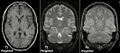
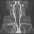
MRI is widely used in hospitals and clinics for medical diagnosis, staging of disease, and follow-up without exposure to ionizing radiation. It is particularly useful for the imaging of the brain, spine, and joints, as well as soft tissues of the musculoskeletal system.
Safety[edit]
MRI is generally safe; it does not involve exposure to ionizing radiation, such as X-rays. However, the presence of strong magnetic fields requires that metal objects are not present in the scanner, and patients with certain types of medical implants, such as pacemakers, may not be suitable candidates for an MRI.
Advancements[edit]
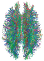
Recent advancements in MRI technology include high-field MRI, functional MRI (fMRI), which measures brain activity by detecting changes associated with blood flow, and real-time MRI, which provides images in real time.
Challenges[edit]
Challenges in MRI technology include reducing scan time, improving image quality, and making MRI accessible in terms of cost and availability.
See also[edit]
This medical article is a stub. You can help WikiMD by expanding the page. |
-
Magnetic resonance imaging
-
Magnetic resonance imaging
-
Magnetic resonance imaging
-
Magnetic resonance imaging
-
 Magnetic resonance imaging
 -
Magnetic resonance imaging
-
Magnetic resonance imaging
-
Magnetic resonance imaging
-
 Magnetic resonance imaging
 -
Magnetic resonance imaging
-
 Magnetic resonance imaging
 -
Magnetic resonance imaging


Medical Disclaimer: WikiMD is for informational purposes only and is not a substitute for professional medical advice. Content may be inaccurate or outdated and should not be used for diagnosis or treatment. Always consult your healthcare provider for medical decisions. Verify information with trusted sources such as CDC.gov and NIH.gov. By using this site, you agree that WikiMD is not liable for any outcomes related to its content. See full disclaimer.
Credits:Most images are courtesy of Wikimedia commons, and templates, categories Wikipedia, licensed under CC BY SA or similar.
Translate page: - East Asian
中文,
日本,
한국어,
South Asian
हिन्दी,
தமிழ்,
తెలుగు,
Urdu,
ಕನ್ನಡ,
Southeast Asian
Indonesian,
Vietnamese,
Thai,
မြန်မာဘာသာ,
বাংলা
European
español,
Deutsch,
français,
Greek,
português do Brasil,
polski,
română,
русский,
Nederlands,
norsk,
svenska,
suomi,
Italian
Middle Eastern & African
عربى,
Turkish,
Persian,
Hebrew,
Afrikaans,
isiZulu,
Kiswahili,
Other
Bulgarian,
Hungarian,
Czech,
Swedish,
മലയാളം,
मराठी,
ਪੰਜਾਬੀ,
ગુજરાતી,
Portuguese,
Ukrainian
