Occipital triangle
Occipital Triangle is an anatomical region located in the posterior aspect of the neck. It is one of the several triangles of the neck and is of clinical importance due to the structures it contains.
Anatomy[edit]
The Occipital Triangle is bounded by the sternocleidomastoid muscle anteriorly, the trapezius muscle posteriorly, and the inferior belly of the omohyoid muscle inferiorly. The floor of the triangle is formed by the splenius capitis, levator scapulae, and the middle and posterior scalene muscles. The roof is formed by the investing layer of the deep cervical fascia.
Contents[edit]
The Occipital Triangle contains several important structures including the Accessory nerve, Cervical plexus, and the Occipital artery.
Accessory Nerve[edit]
The Accessory nerve (Cranial nerve XI) passes diagonally across the triangle from the sternocleidomastoid towards the trapezius.
Cervical Plexus[edit]
The Cervical plexus is a plexus of the anterior rami of the first four cervical spinal nerves which are located from C1 to C4 cervical segments in the neck. They are located laterally to the transverse processes between prevertebral muscles from the medial side and vertebral from lateral side.
Occipital Artery[edit]
The Occipital artery arises from the external carotid artery opposite the facial artery. Its path is below the posterior belly of digastric to the occipital region. This artery supplies blood to the back of the scalp and sterno-mastoid muscles, and deep muscles in the back and neck.
Clinical Significance[edit]
Knowledge of the Occipital Triangle is important in surgeries involving the neck. The Accessory nerve is at risk of injury during surgeries in this region. Damage to this nerve can result in shoulder drop.

This WikiMD article can only be edited by registered and verified editors. You can log in or register.
-
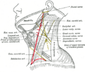
Diagram of the occipital triangle
-
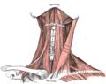
 Muscles of the neck, showing the occipital triangle
 -
 Anatomy of the neck, highlighting the occipital triangle


Medical Disclaimer: WikiMD is for informational purposes only and is not a substitute for professional medical advice. Content may be inaccurate or outdated and should not be used for diagnosis or treatment. Always consult your healthcare provider for medical decisions. Verify information with trusted sources such as CDC.gov and NIH.gov. By using this site, you agree that WikiMD is not liable for any outcomes related to its content. See full disclaimer.
Credits:Most images are courtesy of Wikimedia commons, and templates, categories Wikipedia, licensed under CC BY SA or similar.
Translate page: - East Asian
中文,
日本,
한국어,
South Asian
हिन्दी,
தமிழ்,
తెలుగు,
Urdu,
ಕನ್ನಡ,
Southeast Asian
Indonesian,
Vietnamese,
Thai,
မြန်မာဘာသာ,
বাংলা
European
español,
Deutsch,
français,
Greek,
português do Brasil,
polski,
română,
русский,
Nederlands,
norsk,
svenska,
suomi,
Italian
Middle Eastern & African
عربى,
Turkish,
Persian,
Hebrew,
Afrikaans,
isiZulu,
Kiswahili,
Other
Bulgarian,
Hungarian,
Czech,
Swedish,
മലയാളം,
मराठी,
ਪੰਜਾਬੀ,
ગુજરાતી,
Portuguese,
Ukrainian
