X-ray crystallography
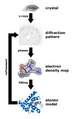
X-ray crystallography is a technique used for determining the atomic and molecular structure of a crystal, in which the crystalline atoms cause a beam of incident X-rays to diffract into many specific directions. By measuring the angles and intensities of these diffracted beams, a crystallographer can produce a three-dimensional picture of the density of electrons within the crystal. From this electron density, the mean positions of the atoms in the crystal can be determined, as well as their chemical bonds, their disorder and various other information.
History[edit]
X-ray crystallography was first used by William Lawrence Bragg and his father, William Henry Bragg, in 1912, following the discovery of X-ray diffraction by Max von Laue in 1912. The Braggs were awarded the Nobel Prize in Physics in 1915 for their work in this field.
Principles[edit]
The basic principle behind X-ray crystallography is Bragg's law, which states that the angles at which a crystal will diffract X-rays are directly related to the size and shape of the unit cell of the crystal. This law is used to determine the distances between planes of atoms within the crystal, which in turn provides information about the crystal's structure.
Applications[edit]
X-ray crystallography has been used in a wide range of fields, including chemistry, physics, materials science, geology, and biology. It has been instrumental in the development of many drugs, as it allows scientists to determine the structure of complex molecules such as proteins and DNA.
See also[edit]
References[edit]

This WikiMD article can only be edited by registered and verified editors. You can log in or register.
X-ray crystallography[edit]
-
 Freezed XRD
 -
 Kepler conjecture 1
 -
 Snowflake
 -
 3D model hydrogen bonds in water
 -
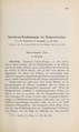 Interferenz-Erscheinungen bei Röntgenstrahlen
 -
 Interferenz-Erscheinungen bei Röntgenstrahlen Tafel II Fig. 5
 -
 Diamond and graphite
 -
 Mars Curiosity Rover 1st X-Ray View
 -
 Penicillin
 -
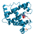 Myoglobin
 -
 X-ray diffraction
 -
 Protein crystal


Medical Disclaimer: WikiMD is for informational purposes only and is not a substitute for professional medical advice. Content may be inaccurate or outdated and should not be used for diagnosis or treatment. Always consult your healthcare provider for medical decisions. Verify information with trusted sources such as CDC.gov and NIH.gov. By using this site, you agree that WikiMD is not liable for any outcomes related to its content. See full disclaimer.
Credits:Most images are courtesy of Wikimedia commons, and templates, categories Wikipedia, licensed under CC BY SA or similar.
Translate page: - East Asian
中文,
日本,
한국어,
South Asian
हिन्दी,
தமிழ்,
తెలుగు,
Urdu,
ಕನ್ನಡ,
Southeast Asian
Indonesian,
Vietnamese,
Thai,
မြန်မာဘာသာ,
বাংলা
European
español,
Deutsch,
français,
Greek,
português do Brasil,
polski,
română,
русский,
Nederlands,
norsk,
svenska,
suomi,
Italian
Middle Eastern & African
عربى,
Turkish,
Persian,
Hebrew,
Afrikaans,
isiZulu,
Kiswahili,
Other
Bulgarian,
Hungarian,
Czech,
Swedish,
മലയാളം,
मराठी,
ਪੰਜਾਬੀ,
ગુજરાતી,
Portuguese,
Ukrainian
