Frozen section procedure
Frozen section procedure is a laboratory technique used in pathology to quickly and effectively diagnose or investigate tissue samples during a surgery. This method involves freezing the tissue specimen, cutting it into very thin sections, and then staining these sections for microscopic examination. The primary advantage of the frozen section procedure is its rapid turnaround time, allowing surgeons to make immediate decisions about the course of treatment based on the pathological findings.
Overview[edit]
The frozen section procedure is typically employed in situations where a quick diagnosis is crucial to the immediate management of the patient. For example, during cancer surgeries, it helps in determining the margins of the tumor—that is, whether the tumor has been completely removed or if additional tissue needs to be excised. It is also used in the diagnosis of suspicious lesions, assessment of lymph nodes during cancer surgery, and in the evaluation of organ transplants.
Procedure[edit]
The process begins with the surgical removal of a tissue sample from the patient. This sample is then rapidly frozen, often by using liquid nitrogen or a cryostat, a device designed specifically for this purpose. Once frozen, the tissue is sliced into thin sections using a microtome within the cryostat. These sections are then placed on slides, stained to highlight different cellular components, and examined under a microscope by a pathologist.
The pathologist assesses the tissue for abnormalities, such as cancer, and communicates the findings to the surgical team. This immediate feedback can be critical in guiding the surgical procedure, such as confirming clear margins in tumor removal or identifying the presence of disease in nearby lymph nodes.
Advantages and Limitations[edit]
The primary advantage of the frozen section procedure is the rapid diagnosis it provides, which is particularly beneficial in the surgical setting. However, it is not without limitations. The quality of the frozen sections can be inferior to that of sections prepared using the more time-consuming paraffin embedding method, potentially making diagnosis more challenging. Additionally, not all tissues are suitable for frozen section analysis, and in some cases, a definitive diagnosis cannot be made, necessitating further testing.
Applications[edit]
The frozen section procedure has a wide range of applications in medical practice, particularly in oncology, where it plays a crucial role in the intraoperative assessment of tumor margins and lymph node involvement. It is also used in the diagnosis of various other conditions, such as infectious diseases and organ transplant rejection.
Conclusion[edit]
The frozen section procedure is a valuable tool in the field of pathology, offering rapid diagnostic capabilities that can significantly impact surgical decisions and patient outcomes. Despite its limitations, the benefits of immediate pathological assessment make it an indispensable technique in many surgical settings.
This medical article is a stub. You can help WikiMD by expanding the page. |
-
 Frozen section procedure
 -
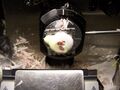
 Putting specimens on chuck
 -
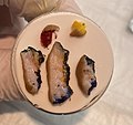
 Covering with embedding medium
 -
 Apply conductor
 -
 Using freeze spray
 -
 Breaking off embedding medium that reach below the chuck's plate
 -
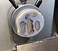 Fastening the chuck on the cryotome and cutting relatively thick sections until the full tissue surfaces of interest are exposed
 -
 Setting the thickness to for example 5 micrometer and sectioning while holding the section down to prevent it from folding onto itself
 -
 Continuing until all the tissue of interest is in the section
 -
 Putting a glass slide on the tissue - version 2
 -
 Time in solutions for frozen sections


Medical Disclaimer: WikiMD is for informational purposes only and is not a substitute for professional medical advice. Content may be inaccurate or outdated and should not be used for diagnosis or treatment. Always consult your healthcare provider for medical decisions. Verify information with trusted sources such as CDC.gov and NIH.gov. By using this site, you agree that WikiMD is not liable for any outcomes related to its content. See full disclaimer.
Credits:Most images are courtesy of Wikimedia commons, and templates, categories Wikipedia, licensed under CC BY SA or similar.
Translate page: - East Asian
中文,
日本,
한국어,
South Asian
हिन्दी,
தமிழ்,
తెలుగు,
Urdu,
ಕನ್ನಡ,
Southeast Asian
Indonesian,
Vietnamese,
Thai,
မြန်မာဘာသာ,
বাংলা
European
español,
Deutsch,
français,
Greek,
português do Brasil,
polski,
română,
русский,
Nederlands,
norsk,
svenska,
suomi,
Italian
Middle Eastern & African
عربى,
Turkish,
Persian,
Hebrew,
Afrikaans,
isiZulu,
Kiswahili,
Other
Bulgarian,
Hungarian,
Czech,
Swedish,
മലയാളം,
मराठी,
ਪੰਜਾਬੀ,
ગુજરાતી,
Portuguese,
Ukrainian
