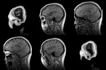
Neuroanatomy
Neuroanatomy refers to the specialized branch of anatomy focusing on the structure and organization of the nervous system. Rooted in both ancient studies and modern science, neuroanatomy has evolved considerably, becoming essential in understanding how the brain and the nervous system function at both microscopic and macroscopic levels.
Introduction
Neuroanatomy delves deep into the structures of the nervous system, from individual neurons to the vast networks that they form. As the intricate structure of the brain and the rest of the nervous system is uncovered, it lays the foundation for various disciplines like neurology, neuropsychology, and neuroscience.
Central vs. Peripheral Nervous System
The nervous system is broadly categorized into two main sections:
- Central Nervous System (CNS): Comprising the brain and the spinal cord, the CNS is the primary control center, processing sensory information and directing responses.
- Peripheral Nervous System (PNS): This system connects the CNS to the rest of the body and includes all neural structures outside of the brain and spinal cord.
Key Structures in Neuroanatomy
The Brain
The brain is a complex organ made up of several regions, each responsible for specific functions:
- Cerebrum: Involved in sensory interpretation, voluntary movements, and cognitive functions.
- Cerebellum: Coordinates muscular activities and maintains posture and balance.
- Brainstem: Controls various involuntary functions such as breathing, heart rate, and blood pressure.
Neurons
Neurons are the building blocks of the nervous system, transmitting electrical and chemical signals. They consist of:
- Dendrites: Receive messages from other neurons.
- Axon: Transmits messages to other neurons or muscles.
- Synapse: The junction where two neurons meet, facilitating signal transmission.
Applications in Medicine and Research
Understanding neuroanatomy has direct implications in fields such as:
- Neurosurgery: Surgical interventions on the nervous system.
- Neuropharmacology: The study of how drugs affect the nervous system.
- Neuroimaging: Techniques like MRI and CT scans used to visualize the structure and function of the nervous system.
| This article is a medical stub. You can help WikiMD by expanding it! | |
|---|---|
| Anatomy and morphology | ||||||||||
|---|---|---|---|---|---|---|---|---|---|---|
|
Transform your life with W8MD's budget GLP-1 injections from $125.
W8MD offers a medical weight loss program to lose weight in Philadelphia. Our physician-supervised medical weight loss provides:
- Most insurances accepted or discounted self-pay rates. We will obtain insurance prior authorizations if needed.
- Generic GLP1 weight loss injections from $125 for the starting dose.
- Also offer prescription weight loss medications including Phentermine, Qsymia, Diethylpropion, Contrave etc.
NYC weight loss doctor appointments
Start your NYC weight loss journey today at our NYC medical weight loss and Philadelphia medical weight loss clinics.
- Call 718-946-5500 to lose weight in NYC or for medical weight loss in Philadelphia 215-676-2334.
- Tags:NYC medical weight loss, Philadelphia lose weight Zepbound NYC, Budget GLP1 weight loss injections, Wegovy Philadelphia, Wegovy NYC, Philadelphia medical weight loss, Brookly weight loss and Wegovy NYC
|
WikiMD's Wellness Encyclopedia |
| Let Food Be Thy Medicine Medicine Thy Food - Hippocrates |
Medical Disclaimer: WikiMD is not a substitute for professional medical advice. The information on WikiMD is provided as an information resource only, may be incorrect, outdated or misleading, and is not to be used or relied on for any diagnostic or treatment purposes. Please consult your health care provider before making any healthcare decisions or for guidance about a specific medical condition. WikiMD expressly disclaims responsibility, and shall have no liability, for any damages, loss, injury, or liability whatsoever suffered as a result of your reliance on the information contained in this site. By visiting this site you agree to the foregoing terms and conditions, which may from time to time be changed or supplemented by WikiMD. If you do not agree to the foregoing terms and conditions, you should not enter or use this site. See full disclaimer.
Credits:Most images are courtesy of Wikimedia commons, and templates, categories Wikipedia, licensed under CC BY SA or similar.
Contributors: Prab R. Tumpati, MD


